CACNG1 Rabbit Polyclonal Antibody

cat.: ER1803-27
| Product Type: | Rabbit polyclonal IgG, primary antibodies |
|---|---|
| Species reactivity: | Human, Mouse, Rat |
| Applications: | WB, IHC-P, IF-Cell, FC |
| Clonality: | Polyclonal |
| Form: | Liquid |
| Storage condition: | Shipped at 4℃. Store at +4℃ short term (1-2 weeks). It is recommended to aliquot into single-use upon delivery. Store at -20℃ long term. |
| Storage buffer: | 1*PBS (pH7.4), 0.2% BSA, 50% Glycerol. Preservative: 0.05% Sodium Azide. |
| Concentration: | 1ug/ul |
| Purification: | Immunogen affinity purified. |
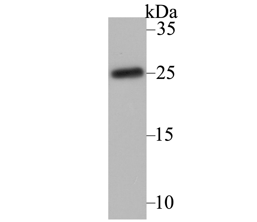
| Molecular weight: | Predicted band size: 25 kDa |
| Isotype: | IgG |
| Immunogen: | Synthetic peptide within mouse CACNG1 aa 13-62 / 223. |
| Positive control: | Human skeletal muscle lysates, human fetal skeletal muscle tissue, mouse skeletal muscle tissue, rat skeletal muscle tissue, SH-SY5Y. |
| Subcellular location: | Cell membrane, sarcolemma. |
| Recommended Dilutions:
WB IHC-P IF-Cell FC |
1:500-1:1,000 1:50-1:200 1:100 1:1,000 |
| Uniprot #: | SwissProt: Q06432 Human | O70578 Mouse | P97707 Rat |
| Alternative names: | CACNG1 CACNLG CACNLG calcium channel neuronal dihydrpyridine sensitive gamma subunit Calcium channel L type gamma polypeptide Calcium channel voltage dependent gamma subunit 1 CCG1_HUMAN Dihydropyridine sensitive L type skeletal muscle calcium channel gamma subunit Dihydropyridine sensitive L type skeletal muscle calcium channel subunit gamma Dihydropyridine-sensitive L-type fb43f02 fi19g08 L type calcium channel gamma polypeptide MGC117703 skeletal muscle calcium channel subunit gamma Voltage dependent calcium channel gamma 1 subunit 2 Voltage-dependent calcium channel gamma-1 subunit wu:fb43f02 zgc:55364 |
Images
|
Fig1: Western blot analysis of CACNG1 on human skeletal muscle lysate using anti-CACNG1 antibody at 1/1,000 dilution. |
|
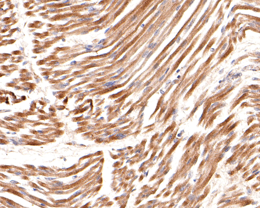
Fig2: Immunohistochemical analysis of paraffin-embedded human fetal skeletal muscle tissue using anti-CACNG1 antibody. Counter stained with hematoxylin. |
|
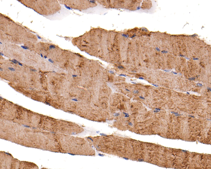
Fig3: Immunohistochemical analysis of paraffin-embedded mouse skeletal muscle tissue using anti-CACNG1 antibody. Counter stained with hematoxylin. |
|
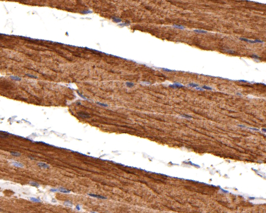
Fig4: Immunohistochemical analysis of paraffin-embedded rat skeletal muscle tissue using anti-CACNG1 antibody. Counter stained with hematoxylin. |

|
Fig5:
Immunocytochemistry analysis of SH-SY5Y cells labeling CACNG1 with Rabbit anti-CACNG1 antibody (ER1803-27) at 1/100 dilution. Cells were fixed in 4% paraformaldehyde for 15 minutes at room temperature, permeabilized with 0.1% Triton X-100 in PBS for 15 minutes at room temperature, then blocked with 1% BSA in 10% negative goat serum for 1 hour at room temperature. Cells were then incubated with Rabbit anti-CACNG1 antibody (ER1803-27) at 1/100 dilution in 1% BSA in PBST overnight at 4 ℃. Goat Anti-Rabbit IgG H&L (iFluor™ 488, HA1121) was used as the secondary antibody at 1/1,000 dilution. PBS instead of the primary antibody was used as the secondary antibody only control. Nuclear DNA was labelled in blue with DAPI. Beta tubulin (HA601187, red) was stained at 1/100 dilution overnight at +4℃. Goat Anti-Mouse IgG H&L (iFluor™ 594, HA1126) was used as the secondary antibody at 1/1,000 dilution. |
|
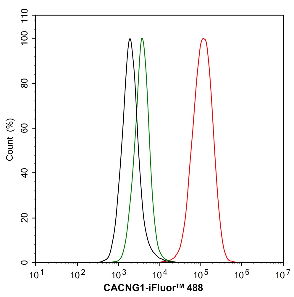
Fig6:
Flow cytometric analysis of SH-SY5Y cells labeling CACNG1. Cells were fixed and permeabilized. Then stained with the primary antibody (ER1803-27, 1/1,000) (red) compared with Rabbit IgG Isotype Control (green). After incubation of the primary antibody at +4℃ for an hour, the cells were stained with a iFluor™ 488 conjugate-Goat anti-Rabbit IgG Secondary antibody (HA1121) at 1/1,000 dilution for 30 minutes at +4℃. Unlabelled sample was used as a control (cells without incubation with primary antibody; black). |
Note: All products are “FOR RESEARCH USE ONLY AND ARE NOT INTENDED FOR DIAGNOSTIC OR THERAPEUTIC USE”.