PPAR gamma Rabbit Polyclonal Antibody

cat.: ER1803-30
| Product Type: | Rabbit polyclonal IgG, primary antibodies |
|---|---|
| Species reactivity: | Human, Mouse, Rat |
| Applications: | WB, IF-Cell, IHC-P, FC |
| Clonality: | Polyclonal |
| Form: | Liquid |
| Storage condition: | Store at +4℃ after thawing. Aliquot store at -20℃. Avoid repeated freeze / thaw cycles. |
| Storage buffer: | 1*PBS (pH7.4), 0.2% BSA, 50% Glycerol. Preservative: 0.05% Sodium Azide. |
| Concentration: | 1ug/ul |
| Purification: | Immunogen affinity purified. |
| Molecular weight: | 57/54/21 kDa |
| Isotype: | IgG |
| Immunogen: | Recombinant protein within human PPAR gamma aa 250-450. |
| Positive control: | PC-12, human liver tissue lysate, A431, A549, JAR, human lung cancer tissue, human colon tissue, mouse uterus tissue. |
| Subcellular location: | Nucleus. Cytoplasm. |
| Recommended Dilutions:
WB IF-Cell IHC-P FC |
1:500 1:100 1:50-1:100 1:50-1:100 |
| Uniprot #: | SwissProt: P37231 Human |
| Alternative names: | CIMT1 GLM1 NR1C3 Nuclear receptor subfamily 1 group C member 3 OTTHUMP00000185032 OTTHUMP00000185036 Peroxisome proliferator activated nuclear receptor gamma variant 1 Peroxisome proliferator activated receptor gamma 1 Peroxisome Proliferator Activated Receptor gamma Peroxisome proliferator-activated receptor gamma PPAR gamma PPAR-gamma PPARG PPARG_HUMAN PPARG1 PPARG2 PPARgamma |
Images
|
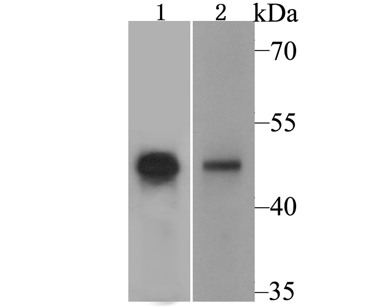
Fig1: Western blot analysis of PPAR gamma on PC-12 cell and human liver tissue lysate using anti-PPAR gamma antibody at 1/500 dilution. |
|
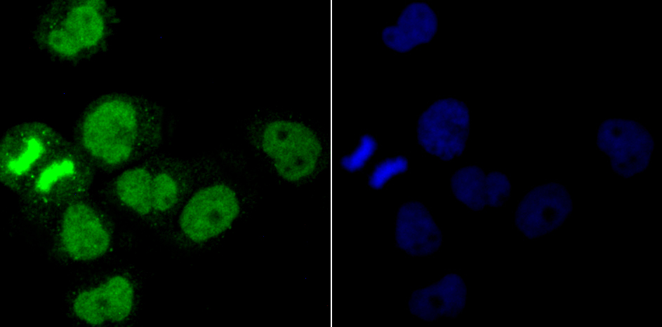
Fig2: ICC staining PPAR gamma in A431 cells (green). The nuclear counter stain is DAPI (blue). Cells were fixed in paraformaldehyde, permeabilised with 0.25% Triton X100/PBS. |
|
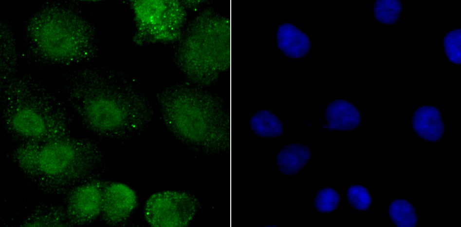
Fig3: ICC staining PPAR gamma in A549 cells (green). The nuclear counter stain is DAPI (blue). Cells were fixed in paraformaldehyde, permeabilised with 0.25% Triton X100/PBS. |
|
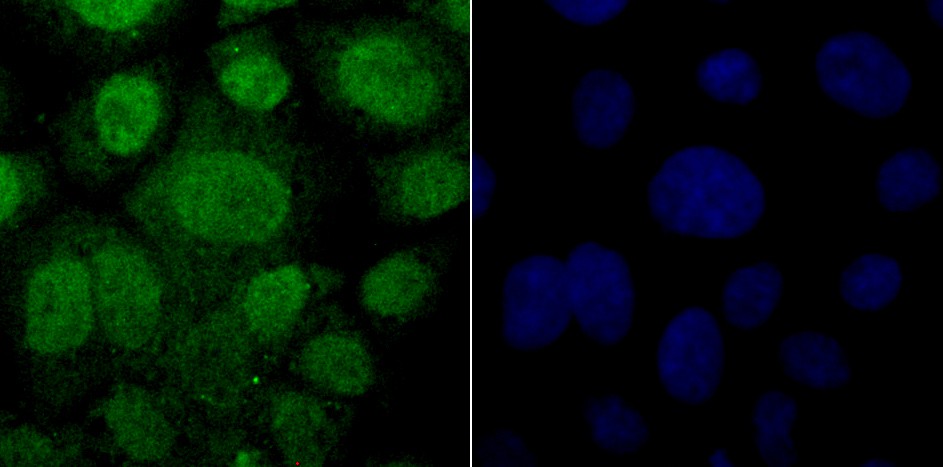
Fig4: ICC staining PPAR gamma in JAR cells (green). The nuclear counter stain is DAPI (blue). Cells were fixed in paraformaldehyde, permeabilised with 0.25% Triton X100/PBS. |
|
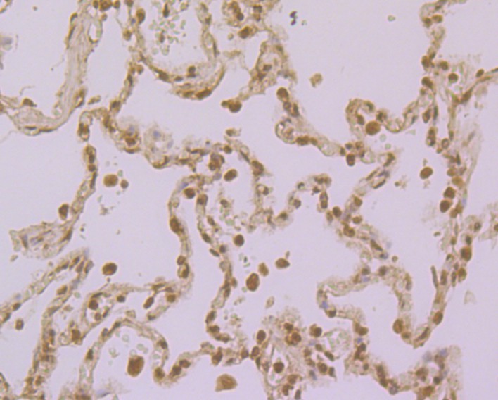
Fig5: Immunohistochemical analysis of paraffin-embedded human lung cancer tissue using anti-PPAR gamma antibody. Counter stained with hematoxylin. |
|
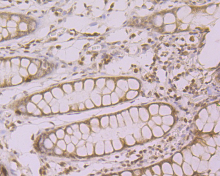
Fig6: Immunohistochemical analysis of paraffin-embedded human colon tissue using anti-PPAR gamma antibody. Counter stained with hematoxylin. |
|
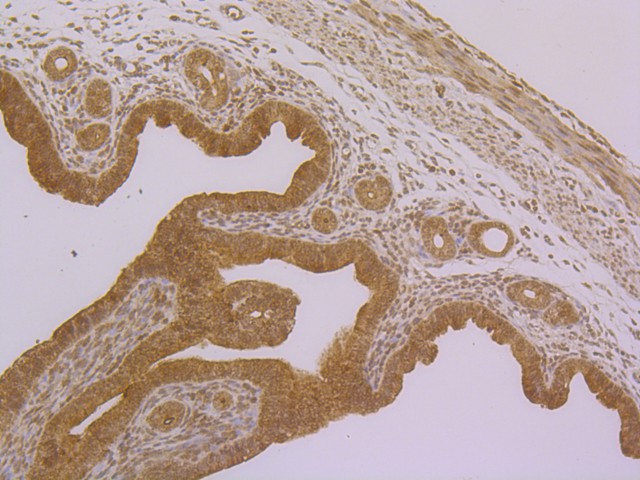
Fig7: Immunohistochemical analysis of paraffin-embedded mouse uterus tissue using anti-PPAR gamma antibody. Counter stained with hematoxylin. |
|
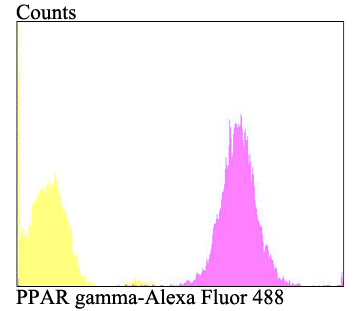
Fig8: Flow cytometric analysis of A431 cells with PPAR gamma antibody at 1/100 dilution (purple) compared with an unlabelled control (cells without incubation with primary antibody; yellow). Alexa Fluor 488-conjugated goat anti-rabbit IgG was used as the secondary antibody. |
Note: All products are “FOR RESEARCH USE ONLY AND ARE NOT INTENDED FOR DIAGNOSTIC OR THERAPEUTIC USE”.