p16INK4a Rabbit Polyclonal Antibody

cat.: ER1803-53
| Product Type: | Rabbit polyclonal IgG, primary antibodies |
|---|---|
| Species reactivity: | Human |
| Applications: | WB, IF-Cell, IHC-P, FC |
| Clonality: | Polyclonal |
| Form: | Liquid |
| Storage condition: | Shipped at 4℃. Store at +4℃ short term (1-2 weeks). It is recommended to aliquot into single-use upon delivery. Store at -20℃ long term. |
| Storage buffer: | 1*PBS (pH7.4), 0.2% BSA, 50% Glycerol. Preservative: 0.05% Sodium Azide. |
| Concentration: | 1ug/ul |
| Purification: | Immunogen affinity purified. |
| Molecular weight: | Predicted band size: 16 kDa |
| Isotype: | IgG |
| Immunogen: | Synthetic peptide within C-terminal human CDKN2A/p16INK4a. |
| Positive control: | HeLa cell lysate, HEK-293 cell lysate, Saos-2 cell lysate, HeLa, human colon cancer tissue, human stomach cancer tissue. |
| Subcellular location: | Nucleus. Cytoplasm. |
| Recommended Dilutions:
WB IF-Cell IHC-P FC |
1:1,000 1:50-1:200 1:50-1:200 1:50-1:100 |
| Uniprot #: | SwissProt: P42771 Human |
| Alternative names: | CCM2 CDK4 inhibitor p16 INK4 CDK4I CDKN2 CDKN2A Cell cycle negative regulator beta CMM2 Cyclin dependent kinase 4 inhibitor A Cyclin dependent kinase inhibitor 2A (melanoma p16 inhibits CDK4) Cyclin Dependent Kinase Inhibitor 2A Cyclin dependent kinase inhibitor 2A isoform 4 Cyclin dependent kinase inhibitor 2A isoforms 1/2/3 Cyclin dependent kinase inhibitor p16 INK4 INK4A MLM MTS1 Multiple tumor suppressor 1 p14 p16 P16INK4 p16INK4a p19 p19Arf TP16 |
Images
|
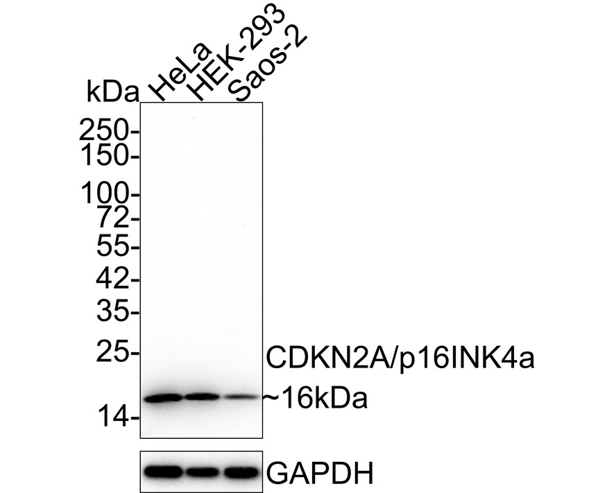
Fig1:
Western blot analysis of p16INK4a on different lysates with Rabbit anti-p16INK4a antibody (ER1803-53) at 1/1,000 dilution. Lane 1: HeLa cell lysate Lane 2: HEK-293 cell lysate Lane 3: Saos-2 cell lysate Lysates/proteins at 20 µg/Lane. Predicted band size: 16 kDa Observed band size: 16 kDa Exposure time: 1 minute; ECL: K1801; 15% SDS-PAGE gel. Proteins were transferred to a PVDF membrane and blocked with 5% NFDM/TBST for 1 hour at room temperature. The primary antibody (ER1803-53) at 1/1,000 dilution was used in 5% NFDM/TBST at 4℃ overnight. Goat Anti-Rabbit IgG - HRP Secondary Antibody (HA1001) at 1:50,000 dilution was used for 1 hour at room temperature. |
|
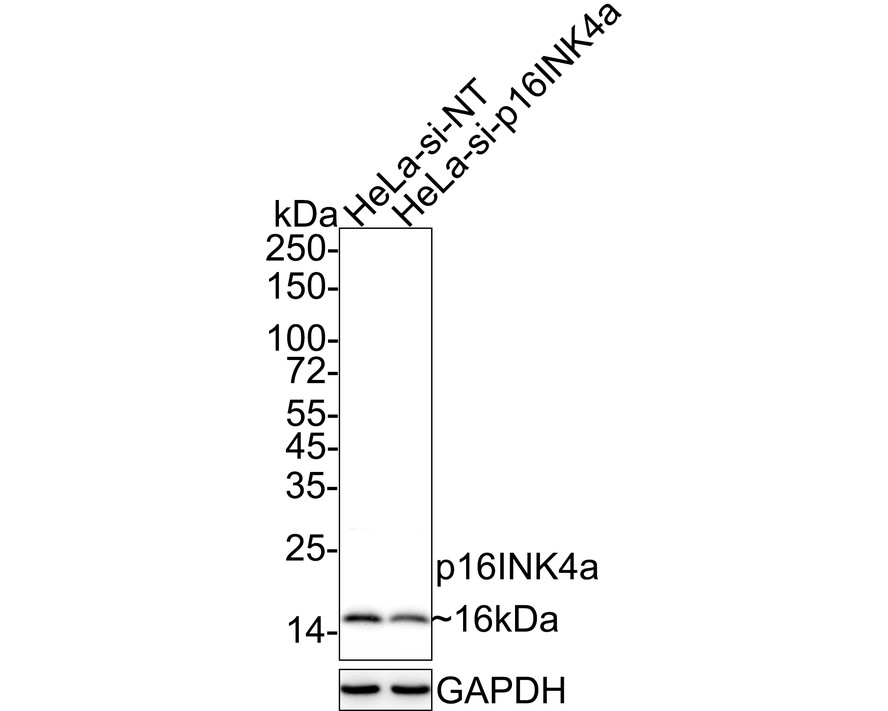
Fig2:
Western blot analysis of p16INK4a on different lysates with Rabbit anti-p16INK4a antibody (ER1803-53) at 1/1,000 dilution. Lane 1: HeLa-si NT cell lysate Lane 2: HeLa-si p16INK4a cell lysate Lysates/proteins at 15 µg/Lane. Predicted band size: 16 kDa Observed band size: 16 kDa Exposure time: 9 seconds; 4-20% SDS-PAGE gel. Proteins were transferred to a PVDF membrane and blocked with 5% NFDM/TBST for 1 hour at room temperature. The primary antibody (ER1803-53) at 1/1,000 dilution was used in 5% NFDM/TBST at 4℃ overnight. Goat Anti-Rabbit IgG - HRP Secondary Antibody (HA1001) at 1/100,000 dilution was used for 1 hour at room temperature. |
|
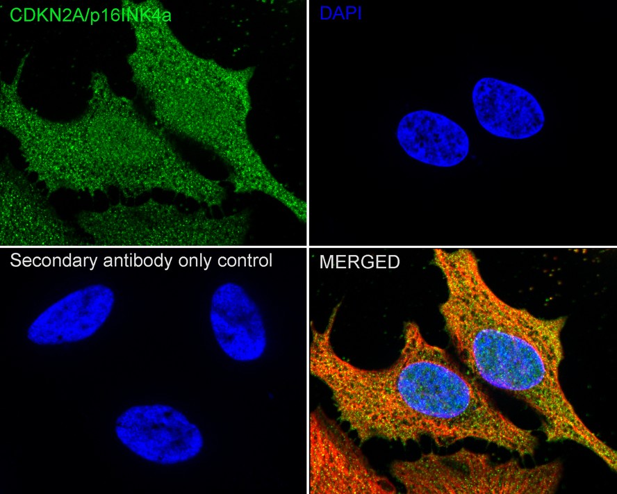
Fig3:
Immunocytochemistry analysis of HeLa cells labeling p16INK4a with Rabbit anti-p16INK4a antibody (ER1803-53) at 1/200 dilution. Cells were fixed in 4% paraformaldehyde for 15 minutes at room temperature, permeabilized with 0.1% Triton X-100 in PBS for 15 minutes at room temperature, then blocked with 1% BSA in 10% negative goat serum for 1 hour at room temperature. Cells were then incubated with Rabbit anti-p16INK4a antibody (ER1803-53) at 1/200 dilution in 1% BSA in PBST overnight at 4 ℃. Goat Anti-Rabbit IgG H&L (iFluor™ 488, HA1121) was used as the secondary antibody at 1/1,000 dilution. PBS instead of the primary antibody was used as the secondary antibody only control. Nuclear DNA was labelled in blue with DAPI. Beta tubulin (HA601187, red) was stained at 1/100 dilution overnight at +4℃. Goat Anti-Mouse IgG H&L (iFluor™ 594, HA1126) was used as the secondary antibody at 1/1,000 dilution. |
|
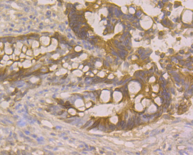
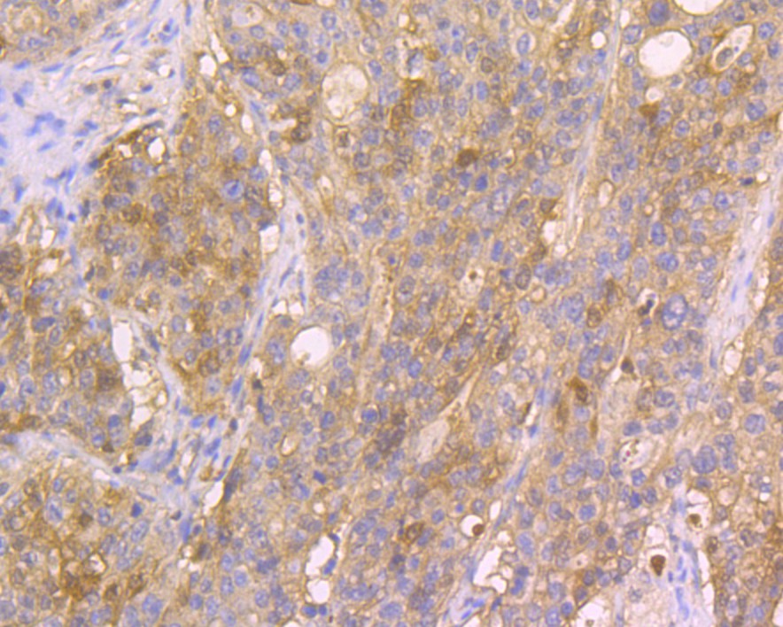
Fig4: Immunohistochemical analysis of paraffin-embedded human colon cancer tissue using anti-CDKN2A/p16INK4a antibody. Counter stained with hematoxylin. The section was pre-treated using heat mediated antigen retrieval with sodium citrate buffer (pH 6.0) (high pressure) for 2 minutes. |
|
Fig5: Immunohistochemical analysis of paraffin-embedded human stomach cancer tissue using anti-CDKN2A/p16INK4a antibody. Counter stained with hematoxylin. The section was pre-treated using heat mediated antigen retrieval with sodium citrate buffer (pH 6.0) (high pressure) for 2 minutes. |
|
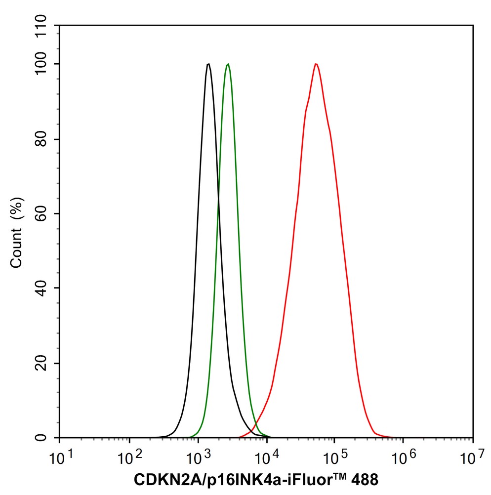
Fig6:
Flow cytometric analysis of HeLa cells labeling p16INK4a. Cells were fixed and permeabilized. Then stained with the primary antibody (ER1803-53, 1/1,000) (red) compared with Rabbit IgG Isotype Control (green). After incubation of the primary antibody at +4℃ for an hour, the cells were stained with a iFluor™ 488 conjugate-Goat anti-Rabbit IgG Secondary antibody (HA1121) at 1/1,000 dilution for 30 minutes at +4℃. Unlabelled sample was used as a control (cells without incubation with primary antibody; black). |
Note: All products are “FOR RESEARCH USE ONLY AND ARE NOT INTENDED FOR DIAGNOSTIC OR THERAPEUTIC USE”.