Acetyl CoA Carboxylase 1 (ACC1) Rabbit Polyclonal Antibody

cat.: ER1803-80
| Product Type: | Rabbit polyclonal IgG, primary antibodies |
|---|---|
| Species reactivity: | Human, Mouse, Rat |
| Applications: | WB, IF-Cell, IHC-P, FC |
| Clonality: | Polyclonal |
| Form: | Liquid |
| Storage condition: | Store at +4℃ after thawing. Aliquot store at -20℃. Avoid repeated freeze / thaw cycles. |
| Storage buffer: | 1*PBS (pH7.4), 0.2% BSA, 50% Glycerol. Preservative: 0.05% Sodium Azide. |
| Concentration: | 1ug/ul |
| Purification: | Immunogen affinity purified. |
| Molecular weight: | 265 kDa |
| Isotype: | IgG |
| Immunogen: | Recombinant protein within Human Acetyl CoA Carboxylase 1 (ACC1) aa 1150-1400. |
| Positive control: | MCF-7, SiHa, rat skeletal muscle tissue, human liver tissue, human kidney tissue, mouse brain tissue. |
| Subcellular location: | Cytoplasm. |
| Recommended Dilutions:
WB IF-Cell IHC-P FC |
1:500 1:50-1:200 1:50-1:200 1:50-1:100 |
| Uniprot #: | SwissProt: Q13085 Human | Q5SWU9 Mouse | P11497 Rat |
| Alternative names: | ACAC ACACA ACACA_HUMAN ACACB ACC alpha ACC ACC beta ACC-alpha ACC1 ACC2 ACCA ACCB Acetyl CoA carboxylase 1 Acetyl CoA carboxylase 2 Acetyl CoA carboxylase alpha Acetyl CoA carboxylase beta Acetyl Coenzyme A carboxylase alpha Acetyl Coenzyme A carboxylase beta Biotin carboxylase COA1 COA2 HACC275 OTTHUMP00000164069 OTTHUMP00000164070 OTTHUMP00000164076 OTTHUMP00000240532 |
Images
|
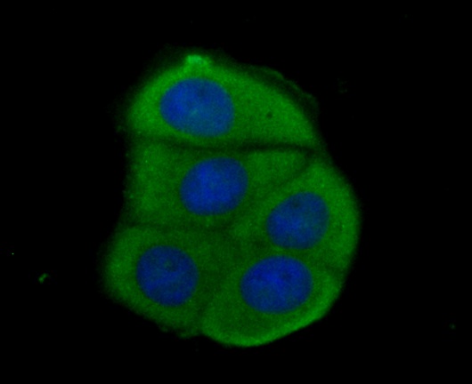
Fig1: ICC staining Acetyl CoA Carboxylase 1 (ACC1) in MCF-7 cells (green). Formalin fixed cells were permeabilized with 0.1% Triton X-100 in TBS for 10 minutes at room temperature and blocked with 1% Blocker BSA for 15 minutes at room temperature. Cells were probed with the antibody (ER1803-80) at a dilution of 1:100 for 1 hour at room temperature, washed with PBS. Alexa Fluorc™ 488 Goat anti-Rabbit IgG was used as the secondary antibody at 1/100 dilution. The nuclear counter stain is DAPI (blue). |
|
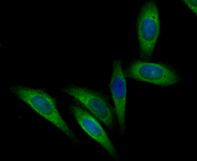
Fig2: ICC staining Acetyl CoA Carboxylase 1 (ACC1) in SiHa cells (green). Formalin fixed cells were permeabilized with 0.1% Triton X-100 in TBS for 10 minutes at room temperature and blocked with 1% Blocker BSA for 15 minutes at room temperature. Cells were probed with the antibody (ER1803-80) at a dilution of 1:200 for 1 hour at room temperature, washed with PBS. Alexa Fluorc™ 488 Goat anti-Rabbit IgG was used as the secondary antibody at 1/100 dilution. The nuclear counter stain is DAPI (blue). |
|
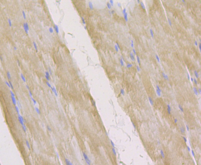
Fig3: Immunohistochemical analysis of paraffin-embedded rat skeletal muscle tissue using anti-Acetyl CoA Carboxylase 1 (ACC1) antibody. The section was pre-treated using heat mediated antigen retrieval with Tris-EDTA buffer (pH 8.0-8.4) for 20 minutes.The tissues were blocked in 5% BSA for 30 minutes at room temperature, washed with ddH2O and PBS, and then probed with the antibody (ER1803-80) at 1/200 dilution, for 30 minutes at room temperature and detected using an HRP conjugated compact polymer system. DAB was used as the chrogen. Counter stained with hematoxylin and mounted with DPX. |
|
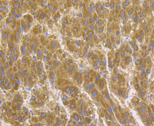
Fig4: Immunohistochemical analysis of paraffin-embedded human liver tissue using anti-Acetyl CoA Carboxylase 1 (ACC1) antibody. The section was pre-treated using heat mediated antigen retrieval with Tris-EDTA buffer (pH 8.0-8.4) for 20 minutes.The tissues were blocked in 5% BSA for 30 minutes at room temperature, washed with ddH2O and PBS, and then probed with the antibody (ER1803-80) at 1/200 dilution, for 30 minutes at room temperature and detected using an HRP conjugated compact polymer system. DAB was used as the chrogen. Counter stained with hematoxylin and mounted with DPX. |
|
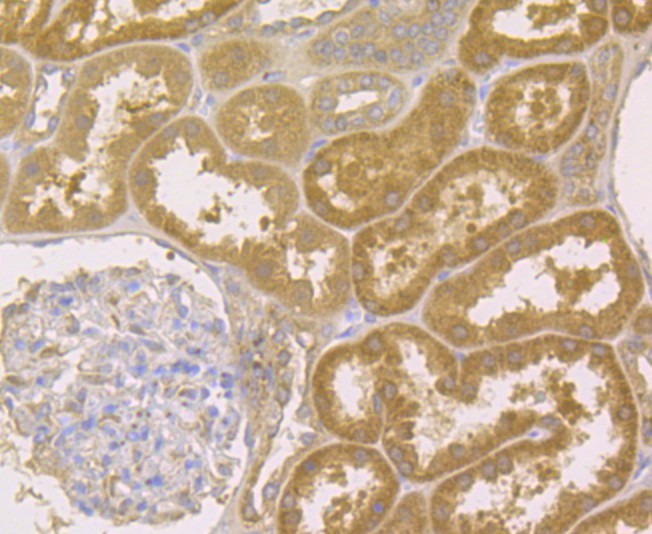
Fig5: Immunohistochemical analysis of paraffin-embedded human kidney tissue using anti-Acetyl CoA Carboxylase 1 (ACC1) antibody. The section was pre-treated using heat mediated antigen retrieval with Tris-EDTA buffer (pH 8.0-8.4) for 20 minutes.The tissues were blocked in 5% BSA for 30 minutes at room temperature, washed with ddH2O and PBS, and then probed with the antibody (ER1803-80) at 1/200 dilution, for 30 minutes at room temperature and detected using an HRP conjugated compact polymer system. DAB was used as the chrogen. Counter stained with hematoxylin and mounted with DPX. |
|
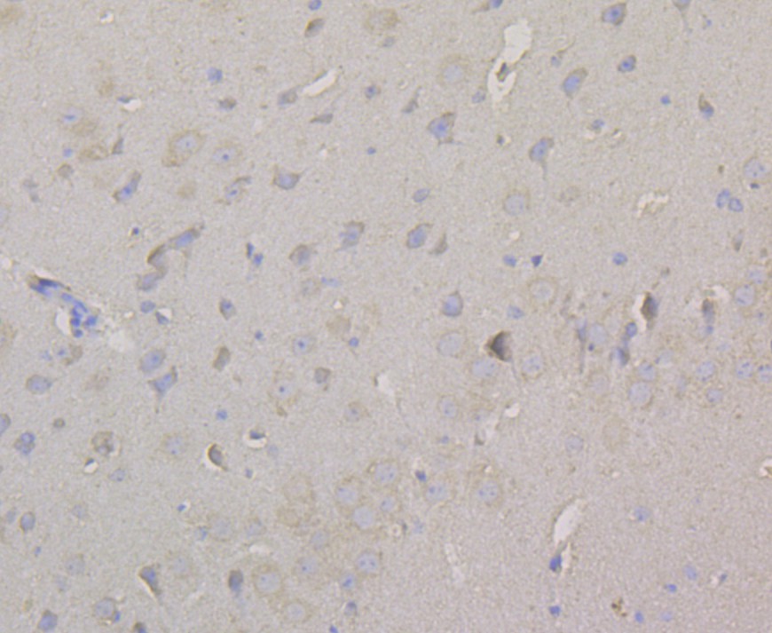
Fig6: Immunohistochemical analysis of paraffin-embedded mouse brain tissue using anti-Acetyl CoA Carboxylase 1 (ACC1) antibody. The section was pre-treated using heat mediated antigen retrieval with Tris-EDTA buffer (pH 8.0-8.4) for 20 minutes.The tissues were blocked in 5% BSA for 30 minutes at room temperature, washed with ddH2O and PBS, and then probed with the antibody (ER1803-80) at 1/200 dilution, for 30 minutes at room temperature and detected using an HRP conjugated compact polymer system. DAB was used as the chrogen. Counter stained with hematoxylin and mounted with DPX. |
Note: All products are “FOR RESEARCH USE ONLY AND ARE NOT INTENDED FOR DIAGNOSTIC OR THERAPEUTIC USE”.