KCNB1 Rabbit Polyclonal Antibody

cat.: ER1901-41
| Product Type: | Rabbit polyclonal IgG, primary antibodies |
|---|---|
| Species reactivity: | Human, Mouse, Rat |
| Applications: | WB, IHC-P, FC |
| Clonality: | Polyclonal |
| Form: | Liquid |
| Storage condition: | Shipped at 4℃. Store at +4℃ short term (1-2 weeks). It is recommended to aliquot into single-use upon delivery. Store at -20℃ long term. |
| Storage buffer: | 1*PBS (pH7.4), 0.2% BSA, 50% Glycerol. Preservative: 0.05% Sodium Azide. |
| Concentration: | 1ug/ul |
| Purification: | Immunogen affinity purified. |
| Molecular weight: | Predicted band size: 96 kDa |
| Isotype: | IgG |
| Immunogen: | Synthetic peptide within rat KCNB1 aa 808-857 / 857. |
| Positive control: | Mouse brain tissue lysate, rat brain tissue lysate, mouse retina tissue lysates, human brain tissue, mouse brain tissue, rat brain tissue, SH-SY5Y. |
| Subcellular location: | Cell membrane, Cell projection, Membrane, Postsynaptic cell membrane, Synapse, Synaptosome. |
| Recommended Dilutions:
WB IHC-P FC |
1:500-1:2,000 1:50-1:200 1:50-1:100 |
| Uniprot #: | SwissProt: Q14721 Human | Q03717 Mouse | P15387 Rat |
| Alternative names: | Delayed rectifier potassium channel 1 Delayed rectifier potassium channel Kv2.1 DRK 1 DRK1 h DRK1 K(+) channel h-DRK1 hDRK 1 hDRK1 KCB 1 KCB1 KCNB1 KCNB1_HUMAN KV2.1 Potassium channel protein DRK1 Potassium voltage gated channel shab related subfamily member 1 Potassium voltage-gated channel subfamily B member 1 Voltage-gated potassium channel subunit Kv2.1 |
Images
|
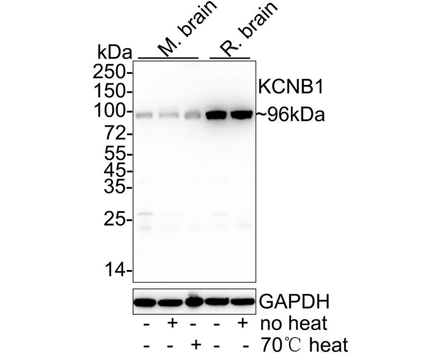
Fig1:
Western blot analysis of KCNB1 on different lysates with Rabbit anti-KCNB1 antibody (ER1901-41) at 1/1,000 dilution. Lane 1: Mouse brain tissue lysate Lane 2: Mouse brain tissue lysate (no heat) Lane 3: Mouse brain tissue lysate (70℃ heat) Lane 4: Rat brain tissue lysate Lane 5: Rat brain tissue lysate (no heat) Notice: no heat means the lysate is not boiled. Lysates/proteins at 40 µg/Lane. Predicted band size: 96 kDa Observed band size: 96 kDa Exposure time: 40 seconds; ECL: K1801; 4-20% SDS-PAGE gel. Proteins were transferred to a PVDF membrane and blocked with 5% NFDM/TBST for 1 hour at room temperature. The primary antibody (ER1901-41) at 1/1,000 dilution was used in 5% NFDM/TBST at room temperature for 2 hours. Goat Anti-Rabbit IgG - HRP Secondary Antibody (HA1001) at 1/50,000 dilution was used for 1 hour at room temperature. |
|
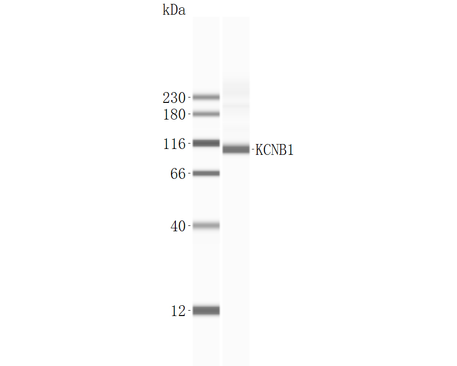
Fig2:
Western blot analysis of KCNB1 on mouse retina tissue lysates with Rabbit anti-KCNB1 antibody (ER1901-41). Predicted band size: 96 kDa Observed band size: 96 kDa |
|
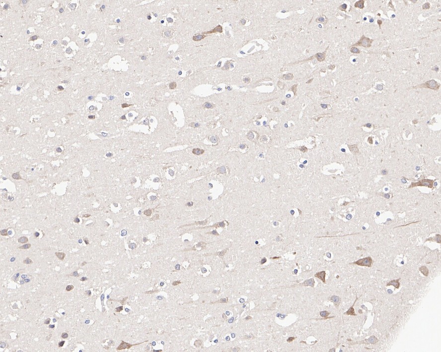
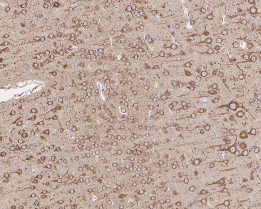
Fig3:
Immunohistochemical analysis of paraffin-embedded human brain tissue with Rabbit anti-KCNB1 antibody (ER1901-41) at 1/200 dilution. The section was pre-treated using heat mediated antigen retrieval with Tris-EDTA buffer (pH 9.0) for 20 minutes. The tissues were blocked in 1% BSA for 20 minutes at room temperature, washed with ddH2O and PBS, and then probed with the primary antibody (ER1901-41) at 1/200 dilution for 1 hour at room temperature. The detection was performed using an HRP conjugated compact polymer system. DAB was used as the chromogen. Tissues were counterstained with hematoxylin and mounted with DPX. |
|
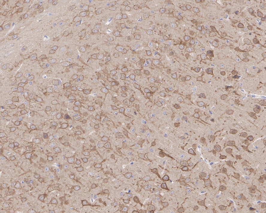
Fig4:
Immunohistochemical analysis of paraffin-embedded mouse brain tissue with Rabbit anti-KCNB1 antibody (ER1901-41) at 1/200 dilution. The section was pre-treated using heat mediated antigen retrieval with Tris-EDTA buffer (pH 9.0) for 20 minutes. The tissues were blocked in 1% BSA for 20 minutes at room temperature, washed with ddH2O and PBS, and then probed with the primary antibody (ER1901-41) at 1/200 dilution for 1 hour at room temperature. The detection was performed using an HRP conjugated compact polymer system. DAB was used as the chromogen. Tissues were counterstained with hematoxylin and mounted with DPX. |
|
Fig5:
Immunohistochemical analysis of paraffin-embedded rat brain tissue with Rabbit anti-KCNB1 antibody (ER1901-41) at 1/200 dilution. The section was pre-treated using heat mediated antigen retrieval with Tris-EDTA buffer (pH 9.0) for 20 minutes. The tissues were blocked in 1% BSA for 20 minutes at room temperature, washed with ddH2O and PBS, and then probed with the primary antibody (ER1901-41) at 1/200 dilution for 1 hour at room temperature. The detection was performed using an HRP conjugated compact polymer system. DAB was used as the chromogen. Tissues were counterstained with hematoxylin and mounted with DPX. |
|
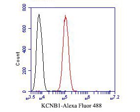
Fig6: Flow cytometric analysis of KCNB1 was done on SH-SY5Y cells. The cells were fixed, permeabilized and stained with the primary antibody (ER1901-41, 1/50) (red). After incubation of the primary antibody at room temperature for an hour, the cells were stained with a Alexa Fluor 488-conjugated Goat anti-Rabbit IgG Secondary antibody at 1/1000 dilution for 30 minutes.Unlabelled sample was used as a control (cells without incubation with primary antibody; black). |
Note: All products are “FOR RESEARCH USE ONLY AND ARE NOT INTENDED FOR DIAGNOSTIC OR THERAPEUTIC USE”.