Syntrophin alpha 1 Rabbit Polyclonal Antibody

cat.: ER1901-45
| Product Type: | Rabbit polyclonal IgG, primary antibodies |
|---|---|
| Species reactivity: | Human, Mouse, Rat |
| Applications: | WB, IF-Cell, IHC-P, FC |
| Clonality: | Polyclonal |
| Form: | Liquid |
| Storage condition: | Store at +4℃ after thawing. Aliquot store at -20℃. Avoid repeated freeze / thaw cycles. |
| Storage buffer: | 1*PBS (pH7.4), 0.2% BSA, 50% Glycerol. Preservative: 0.05% Sodium Azide. |
| Concentration: | 1ug/ul |
| Purification: | Immunogen affinity purified. |
| Molecular weight: | Predicted band size: 54 kDa |
| Isotype: | IgG |
| Immunogen: | Synthetic peptide within mouse Syntrophin alpha 1 aa 174-223 / 503. |
| Positive control: | Mouse skeletal muscle tissue lysate, rat skeletal muscle tissue lysate, SW620, human kidney tissue, mouse skeletal muscle tissue, SiHa. |
| Subcellular location: | Cell junction, Cell membrane, Cytoplasm, Cytoskeleton, Membrane. |
| Recommended Dilutions:
WB IHC-P IF-Cell FC |
1:500-1:2,000 1:50-1:100 1:50-1:100 1:50-1:100 |
| Uniprot #: | SwissProt: Q13424 Human | Q61234 Mouse |
| Alternative names: | 59 kDa dystrophin-associated protein A1 acidic component 1 Alpha-1-syntrophin Pro-TGF-alpha cytoplasmic domain-interacting protein 1 SNT1 SNT2 SNT3 Snta1 SNTA1_HUMAN SNTB1 SNTB2 Syntrophin alpha 1 Syntrophin beta 1 Syntrophin beta 2 Syntrophin-1 TACIP1 |
Images
|
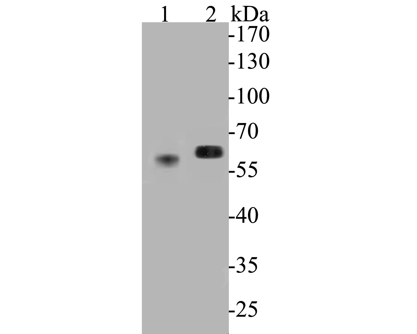
Fig1:
Western blot analysis of Syntrophin alpha 1 on different lysates. Proteins were transferred to a PVDF membrane and blocked with 5% BSA in PBS for 1 hour at room temperature. The primary antibody (ER1901-45, 1/500) was used in 5% BSA at room temperature for 2 hours. Goat Anti-Rabbit IgG - HRP Secondary Antibody (HA1001) at 1:5,000 dilution was used for 1 hour at room temperature. Positive control: Lane 1: mouse skeletal muscle tissue lysate Lane 2: rat skeletal muscle tissue lysate |
|
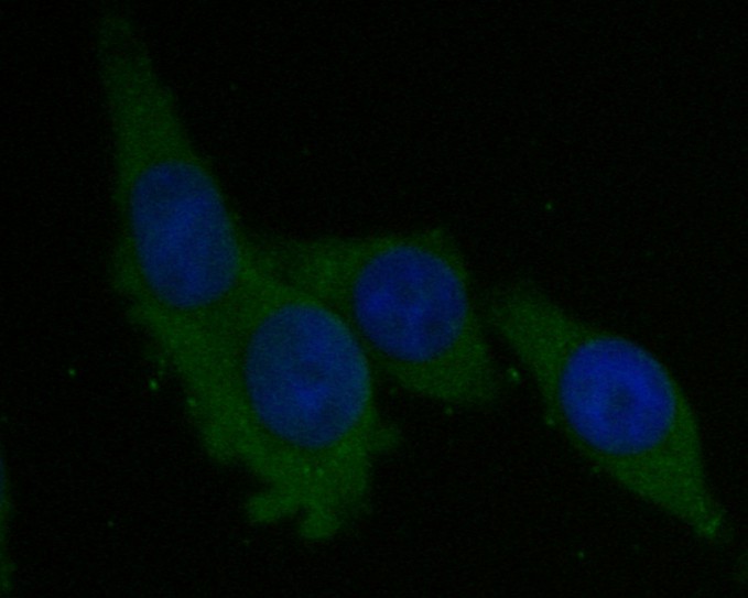
Fig2: ICC staining of Syntrophin alpha 1 in SW620 cells (green). Formalin fixed cells were permeabilized with 0.1% Triton X-100 in TBS for 10 minutes at room temperature and blocked with 1% Blocker BSA for 15 minutes at room temperature. Cells were probed with the primary antibody (ER1901-45, 1/50) for 1 hour at room temperature, washed with PBS. Alexa Fluor®488 Goat anti-Rabbit IgG was used as the secondary antibody at 1/1,000 dilution. The nuclear counter stain is DAPI (blue). |
|
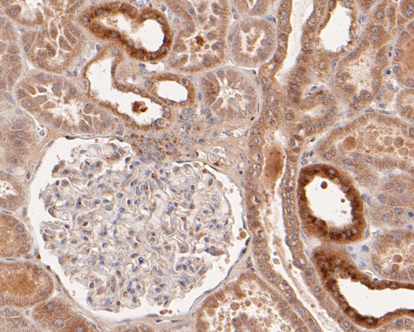
Fig3: Immunohistochemical analysis of paraffin-embedded human kidney tissue using anti-Syntrophin alpha 1 antibody. The section was pre-treated using heat mediated antigen retrieval with Tris-EDTA buffer (pH 8.0-8.4) for 20 minutes.The tissues were blocked in 5% BSA for 30 minutes at room temperature, washed with ddH2O and PBS, and then probed with the primary antibody (ER1901-45, 1/50) for 30 minutes at room temperature. The detection was performed using an HRP conjugated compact polymer system. DAB was used as the chromogen. Tissues were counterstained with hematoxylin and mounted with DPX. |
|
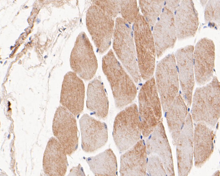
Fig4: Immunohistochemical analysis of paraffin-embedded mouse skeletal muscle tissue using anti-Syntrophin alpha 1 antibody. The section was pre-treated using heat mediated antigen retrieval with Tris-EDTA buffer (pH 8.0-8.4) for 20 minutes.The tissues were blocked in 5% BSA for 30 minutes at room temperature, washed with ddH2O and PBS, and then probed with the primary antibody (ER1901-45, 1/50) for 30 minutes at room temperature. The detection was performed using an HRP conjugated compact polymer system. DAB was used as the chromogen. Tissues were counterstained with hematoxylin and mounted with DPX. |
|
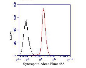
Fig5: Flow cytometric analysis of Syntrophin alpha 1 was done on SiHa cells. The cells were fixed, permeabilized and stained with the primary antibody (ER1901-45, 1/50) (red). After incubation of the primary antibody at room temperature for an hour, the cells were stained with a Alexa Fluor 488-conjugated Goat anti-Rabbit IgG Secondary antibody at 1/1000 dilution for 30 minutes.Unlabelled sample was used as a control (cells without incubation with primary antibody; black). |
Note: All products are “FOR RESEARCH USE ONLY AND ARE NOT INTENDED FOR DIAGNOSTIC OR THERAPEUTIC USE”.