GAPDH Rabbit Polyclonal Antibody

cat.: ER1901-65
| Product Type: | Rabbit polyclonal IgG, primary antibodies |
|---|---|
| Species reactivity: | Human, Mouse, Rat |
| Applications: | WB, IHC-P, IF-Cell, FC |
| Clonality: | Polyclonal |
| Form: | Liquid |
| Storage condition: | Shipped at 4℃. Store at +4℃ short term (1-2 weeks). It is recommended to aliquot into single-use upon delivery. Store at -20℃ long term. |
| Storage buffer: | 1*PBS (pH7.4), 0.2% BSA, 50% Glycerol. Preservative: 0.05% Sodium Azide. |
| Concentration: | 1ug/ul |
| Purification: | Protein A affinity purified. |
| Molecular weight: | 36 kDa |
| Isotype: | IgG |
| Immunogen: | Recombinant protein within Human GAPDH aa 15-171. |
| Positive control: | PC-12 cell lysate, Hela cell lysate, mouse ovary tissue lysate, human placenta tissue lysate, rat brain tissue lysate, NCCIT cell lysate, A549, LoVo, MCF-7, rat kidney tissue, human colon cancer tissue, human spleen tissue, mouse testis tissue. |
| Subcellular location: | Cytoplasm, Nucleus, Membrane. |
| Recommended Dilutions:
WB IF-Cell IHC-P FC |
1:500-1:2,000 1:50-1:200 1:50-1:200 1:50-1:100 |
| Uniprot #: | SwissProt: P04406 Human | P16858 Mouse | P04797 Rat |
| Alternative names: | 38 kDa BFA-dependent ADP-ribosylation substrate aging associated gene 9 protein Aging-associated gene 9 protein BARS-38 cb609 EC 1.2.1.12 Epididymis secretory sperm binding protein Li 162eP G3P_HUMAN G3PD G3PDH GAPD GAPDH Glyceraldehyde 3 phosphate dehydrogenase Glyceraldehyde-3-phosphate dehydrogenase HEL-S-162eP KNC-NDS6 MGC102544 MGC102546 MGC103190 MGC103191 MGC105239 MGC127711 MGC88685 OCAS, p38 component OCT1 coactivator in S phase, 38-KD component peptidyl cysteine S nitrosylase GAPDH Peptidyl-cysteine S-nitrosylase GAPDH wu:fb33a10 |
Images
|
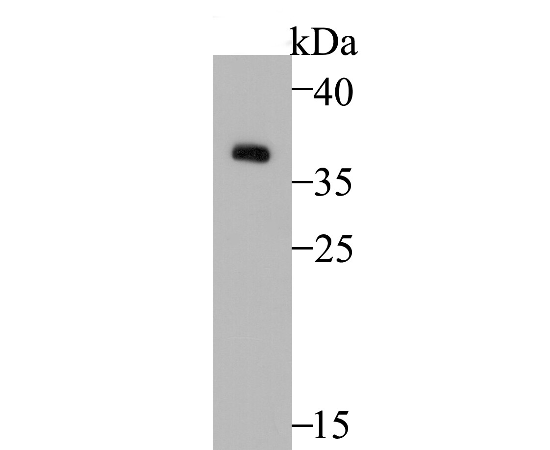
Fig1: Western blot analysis of GAPDH on PC-12 cell lysate. Proteins were transferred to a PVDF membrane and blocked with 5% BSA in PBS for 1 hour at room temperature. The primary antibody was used at a 1:2,000 dilution in 5% BSA at room temperature for 2 hours. Goat Anti-Rabbit IgG - HRP Secondary Antibody (HA1001) at 1:5,000 dilution was used for 1 hour at room temperature. |
|
Fig2:
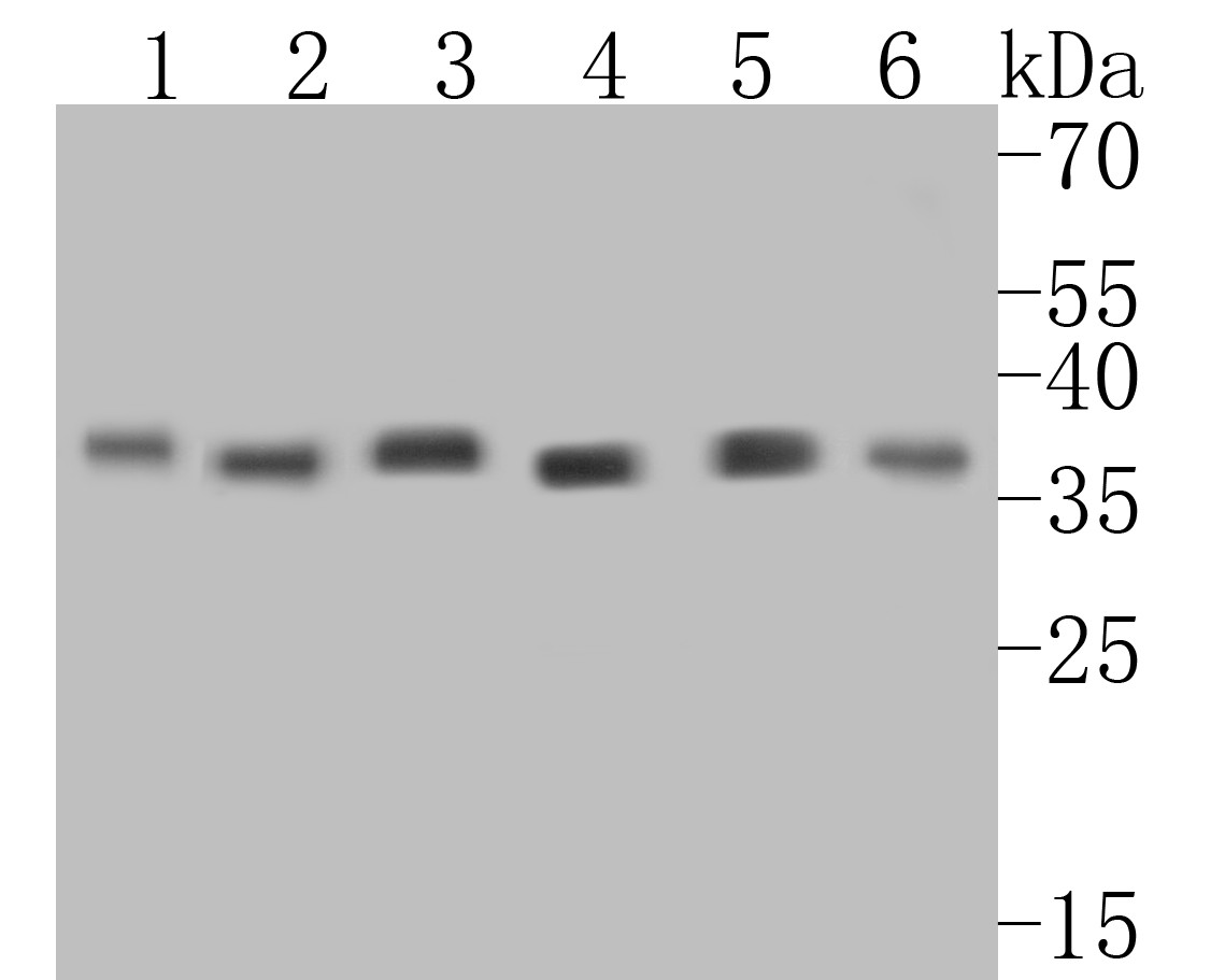
Western blot analysis of GAPDH on different lysates. Proteins were transferred to a PVDF membrane and blocked with 5% BSA in PBS for 1 hour at room temperature. The primary antibody (ER1901-65, 1/500) was used in 5% BSA at room temperature for 2 hours. Goat Anti-Rabbit IgG - HRP Secondary Antibody (HA1001) at 1:5,000 dilution was used for 1 hour at room temperature. Positive control: Lane 1: PC-12 cell lysate Lane 2: Hela cell lysate Lane 3: Mouse ovary tissue lysate Lane 4: Human placenta tissue lysate Lane 5: Rat brain tissue lysate Lane 6: NCCIT cell lysate |
|
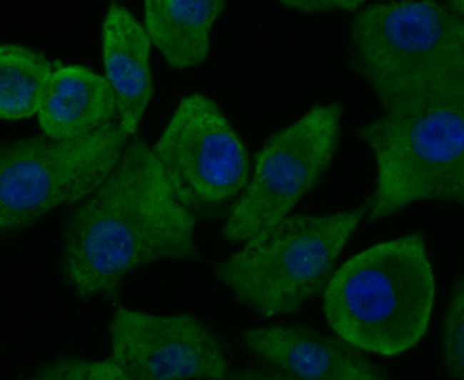
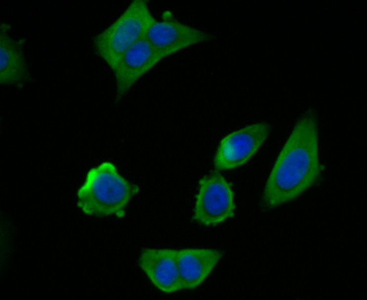
Fig3: ICC staining of GAPDH in A549 cells (green). Formalin fixed cells were permeabilized with 0.1% Triton X-100 in TBS for 10 minutes at room temperature and blocked with 1% Blocker BSA for 15 minutes at room temperature. Cells were probed with the antibody (ER1901-65) at a dilution of 1:50 for 1 hour at room temperature, washed with PBS. Alexa Fluor®488 Goat anti-Rabbit IgG was used as the secondary antibody at 1/100 dilution. The nuclear counter stain is DAPI (blue). |
|
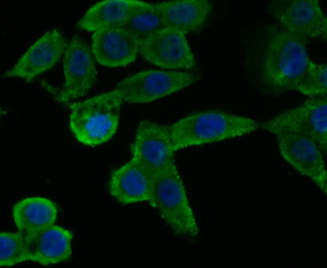
Fig4: ICC staining of GAPDH in LoVo cells (green). Formalin fixed cells were permeabilized with 0.1% Triton X-100 in TBS for 10 minutes at room temperature and blocked with 1% Blocker BSA for 15 minutes at room temperature. Cells were probed with the antibody (ER1901-65) at a dilution of 1:50 for 1 hour at room temperature, washed with PBS. Alexa Fluor®488 Goat anti-Rabbit IgG was used as the secondary antibody at 1/100 dilution. The nuclear counter stain is DAPI (blue). |
|
Fig5: ICC staining of GAPDH in MCF-7 cells (green). Formalin fixed cells were permeabilized with 0.1% Triton X-100 in TBS for 10 minutes at room temperature and blocked with 1% Blocker BSA for 15 minutes at room temperature. Cells were probed with the antibody (ER1901-65) at a dilution of 1:100 for 1 hour at room temperature, washed with PBS. Alexa Fluor®488 Goat anti-Rabbit IgG was used as the secondary antibody at 1/100 dilution. The nuclear counter stain is DAPI (blue). |
|
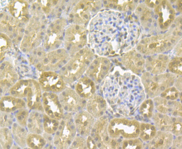
Fig6: Immunohistochemical analysis of paraffin-embedded rat kidney tissue using anti-GAPDH antibody. The section was pre-treated using heat mediated antigen retrieval with sodium citrate buffer (pH 6.0) for 20 minutes. The tissues were blocked in 5% BSA for 30 minutes at room temperature, washed with ddH2O and PBS, and then probed with the antibody (ER1901-65) at 1/50 dilution, for 30 minutes at room temperature and detected using an HRP conjugated compact polymer system. DAB was used as the chromogen. Counter stained with hematoxylin and mounted with DPX. |
|
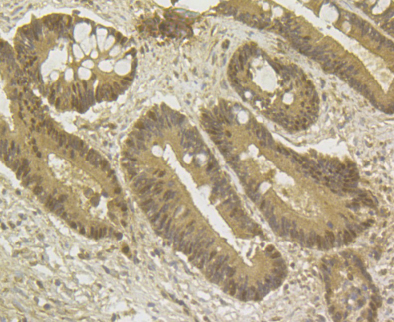
Fig7: Immunohistochemical analysis of paraffin-embedded human colon cancer tissue using anti-GAPDH antibody. The section was pre-treated using heat mediated antigen retrieval with sodium citrate buffer (pH 6.0) for 20 minutes. The tissues were blocked in 5% BSA for 30 minutes at room temperature, washed with ddH2O and PBS, and then probed with the antibody (ER1901-65) at 1/50 dilution, for 30 minutes at room temperature and detected using an HRP conjugated compact polymer system. DAB was used as the chromogen. Counter stained with hematoxylin and mounted with DPX. |
|
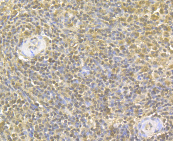
Fig8: Immunohistochemical analysis of paraffin-embedded human spleen tissue using anti-GAPDH antibody. The section was pre-treated using heat mediated antigen retrieval with sodium citrate buffer (pH 6.0) for 20 minutes. The tissues were blocked in 5% BSA for 30 minutes at room temperature, washed with ddH2O and PBS, and then probed with the antibody (ER1901-65) at 1/50 dilution, for 30 minutes at room temperature and detected using an HRP conjugated compact polymer system. DAB was used as the chromogen. Counter stained with hematoxylin and mounted with DPX. |
|
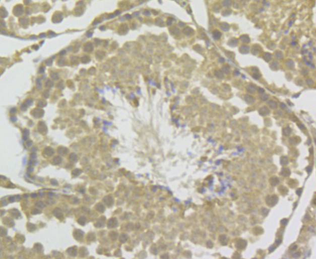
Fig9: Immunohistochemical analysis of paraffin-embedded mouse testis tissue using anti-GAPDH antibody. The section was pre-treated using heat mediated antigen retrieval with sodium citrate buffer (pH 6.0) for 20 minutes. The tissues were blocked in 5% BSA for 30 minutes at room temperature, washed with ddH2O and PBS, and then probed with the antibody (ER1901-65) at 1/50 dilution, for 30 minutes at room temperature and detected using an HRP conjugated compact polymer system. DAB was used as the chromogen. Counter stained with hematoxylin and mounted with DPX. |
|
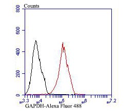
Fig10: Flow cytometric analysis of GAPDH was done on MCF-7 cells. The cells were fixed, permeabilized and stained with GAPDH antibody at 1/50 dilution (red) compared with an unlabelled control (cells without incubation with primary antibody; black). After incubation of the primary antibody on room temperature for an hour, the cells was stained with a Alexa Fluor 488-conjugated goat anti-rabbit IgG Secondary antibody at 1/500 dilution for 30 minutes. |
Note: All products are “FOR RESEARCH USE ONLY AND ARE NOT INTENDED FOR DIAGNOSTIC OR THERAPEUTIC USE”.