NAPSIN A Rabbit Polyclonal Antibody

cat.: ER1901-84
| Product Type: | Rabbit polyclonal IgG, primary antibodies |
|---|---|
| Species reactivity: | Human |
| Applications: | WB, IF-Cell, IHC-P, FC |
| Clonality: | Polyclonal |
| Form: | Liquid |
| Storage condition: | Store at +4℃ after thawing. Aliquot store at -20℃. Avoid repeated freeze / thaw cycles. |
| Storage buffer: | 1*PBS (pH7.4), 0.2% BSA, 50% Glycerol. Preservative: 0.05% Sodium Azide. |
| Concentration: | 1ug/ul |
| Purification: | Immunogen affinity purified. |
| Molecular weight: | Predicted band size 11/30/45 kDa. |
| Isotype: | IgG |
| Immunogen: | Recombinant protein within Human NAPSIN A aa 44-183 / 420. |
| Positive control: | Human lung tissue, 293T, human lung cancer tissue, human kidney tissue. |
| Subcellular location: | Secreted. |
| Recommended Dilutions:
WB IF-Cell IHC-P FC |
1:500-1:1,000 1:50-1:200 1:50-1:200 1:50-1:100 |
| Uniprot #: | SwissProt: O96009 Human |
| Alternative names: | Asp 4 ASP4 Aspartyl protease 4 KAP Kdap Kidney derived aspartic protease like protein NAP1 NAPA Napsa NAPSA_HUMAN Napsin 1 napsin A aspartic peptidase Napsin A precursor Napsin-1 Napsin-A Pronapsin A SNAPA TA01/TA02 |
Images
|
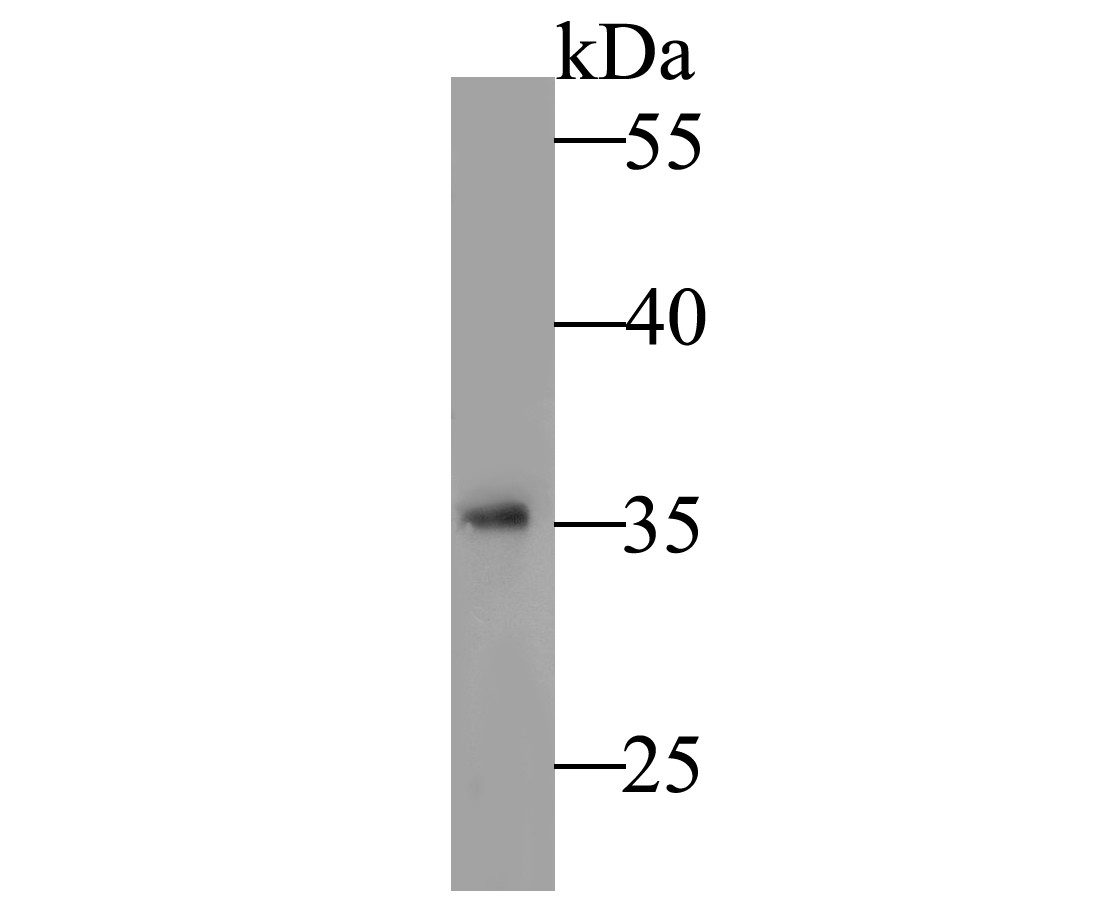
Fig1: Western blot analysis of NAPSIN A on human lung tissue lysate. Proteins were transferred to a PVDF membrane and blocked with 5% BSA in PBS for 1 hour at room temperature. The primary antibody was used at a 1:500 dilution in 5% BSA at room temperature for 2 hours. Goat Anti-Rabbit IgG - HRP Secondary Antibody (HA1001) at 1:5,000 dilution was used for 1 hour at room temperature. |
|
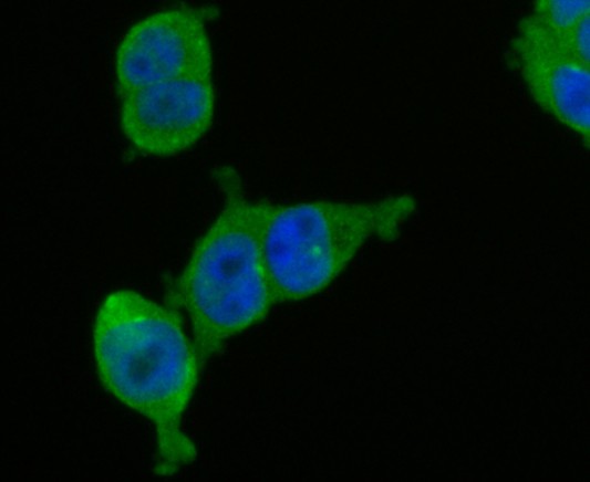
Fig2: ICC staining of NAPSIN A in 293T cells (green). Formalin fixed cells were permeabilized with 0.1% Triton X-100 in TBS for 10 minutes at room temperature and blocked with 1% Blocker BSA for 15 minutes at room temperature. Cells were probed with the primary antibody (ER1901-84, 1/100 dilution) for 1 hour at room temperature, washed with PBS. Alexa Fluor®488 Goat anti-Rabbit IgG was used as the secondary antibody at 1/100 dilution. The nuclear counter stain is DAPI (blue). |
|
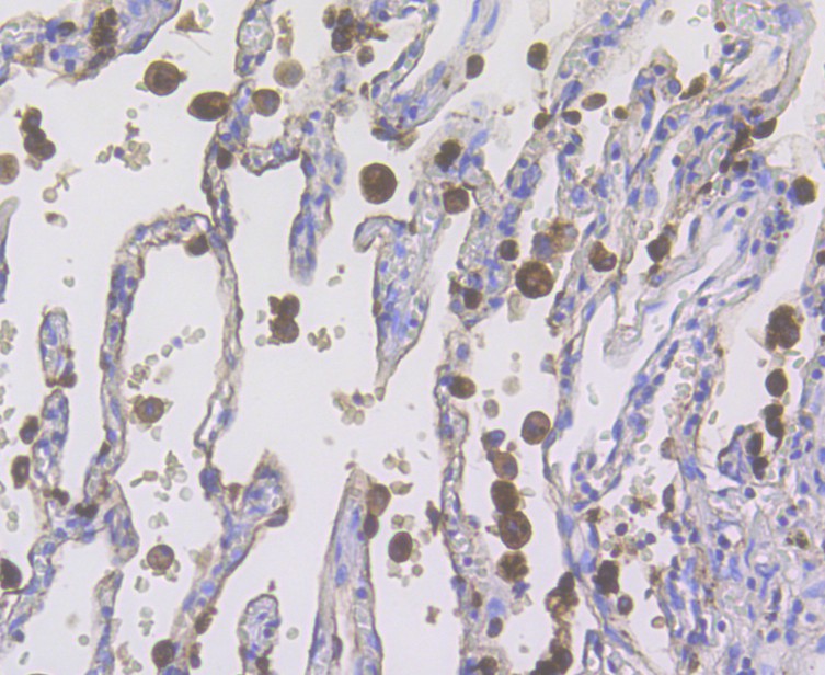
Fig3: Immunohistochemical analysis of paraffin-embedded human lung cancer tissue using anti-NAPSIN A antibody. The section was pre-treated using heat mediated antigen retrieval with Tris-EDTA buffer (pH 8.0-8.4) for 20 minutes.The tissues were blocked in 5% BSA for 30 minutes at room temperature, washed with ddH2O and PBS, and then probed with the primary antibody (ER1901-84, 1/200 dilution) for 30 minutes at room temperature and detected using an HRP conjugated compact polymer system. DAB was used as the chromogen. Counter stained with hematoxylin and mounted with DPX. |
|
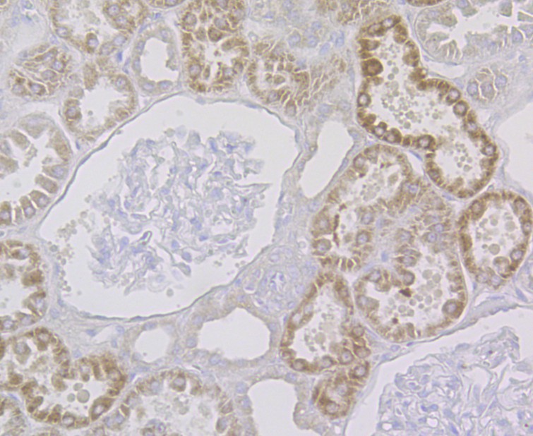
Fig4: Immunohistochemical analysis of paraffin-embedded human kidney tissue using anti-NAPSIN A antibody. The section was pre-treated using heat mediated antigen retrieval with Tris-EDTA buffer (pH 8.0-8.4) for 20 minutes.The tissues were blocked in 5% BSA for 30 minutes at room temperature, washed with ddH2O and PBS, and then probed with the primary antibody (ER1901-84, 1/200 dilution) for 30 minutes at room temperature and detected using an HRP conjugated compact polymer system. DAB was used as the chromogen. Counter stained with hematoxylin and mounted with DPX. |
|
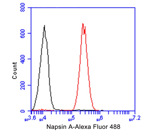
Fig5: Flow cytometric analysis of NAPSIN A was done on A549 cells. The cells were fixed, permeabilized and stained with the primary antibody (ER1901-84, 1/100) (red). After incubation of the primary antibody at room temperature for an hour, the cells were stained with a Alexa Fluor 488-conjugated goat anti-rabbit IgG Secondary antibody at 1/500 dilution for 30 minutes.Unlabelled sample was used as a control (cells without incubation with primary antibody; black). |
Note: All products are “FOR RESEARCH USE ONLY AND ARE NOT INTENDED FOR DIAGNOSTIC OR THERAPEUTIC USE”.