CNGA4 Rabbit Polyclonal Antibody

cat.: ER1901-88
| Product Type: | Rabbit polyclonal IgG, primary antibodies |
|---|---|
| Species reactivity: | Human, Mouse, Rat |
| Applications: | WB, IF-Cell, FC |
| Clonality: | Polyclonal |
| Form: | Liquid |
| Storage condition: | Shipped at 4℃. Store at +4℃ short term (1-2 weeks). It is recommended to aliquot into single-use upon delivery. Store at -20℃ long term. |
| Storage buffer: | 1*PBS (pH7.4), 0.2% BSA, 50% Glycerol. Preservative: 0.05% Sodium Azide. |
| Concentration: | 1ug/ul |
| Purification: | Immunogen affinity purified. |
| Molecular weight: | Predicted band size 65 kDa. |
| Isotype: | IgG |
| Immunogen: | Synthetic peptide within rat CNGA4 aa 1-50 / 575. |
| Positive control: | A549 cell lysate, A431 cell lysate, human brain tissue lysate, SHSY5Y cell lysate, N2A, SHSY5Y, Hela. |
| Subcellular location: | Membrane. |
| Recommended Dilutions:
WB IF-Cell FC |
1:500-1:1000 1:50-1:200 1:50-1:100 |
| Uniprot #: | SwissProt: Q8IV77 Human | Q3UW12 Mouse | Q64359 Rat |
| Alternative names: | Cyclic nucleotide-gated cation channel alpha-4 Cyclic nucleotide-gated channel alpha-4 CNG channel alpha-4 CNG-4 CNG4 Cnga4 Cyclic nucleotide-gated olfactory channel subunit OCNC2 CNG4 |
Images
|
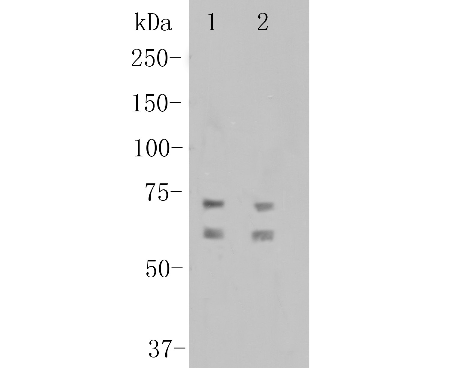
Fig1:
Western blot analysis of CNGA4 on different lysates. Proteins were transferred to a PVDF membrane and blocked with 5% BSA in PBS for 1 hour at room temperature. The primary antibody (ER1901-88, 1/500) was used in 5% BSA at room temperature for 2 hours. Goat Anti-Rabbit IgG - HRP Secondary Antibody (HA1001) at 1:5,000 dilution was used for 1 hour at room temperature. Positive control: Lane 1: A549 cell lysate Lane 2: A431 cell lysate |
|
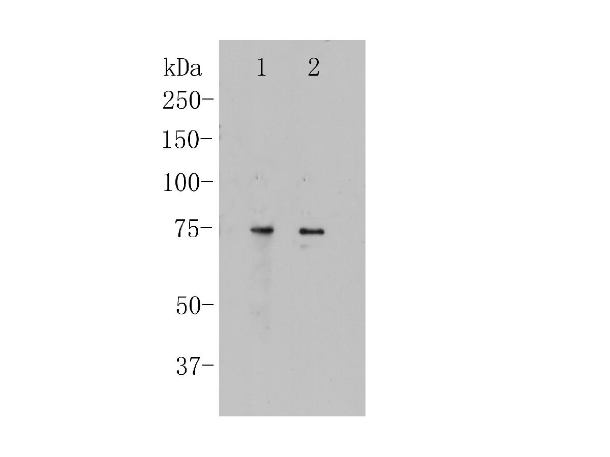
Fig2:
Western blot analysis of CNGA4 on different lysates. Proteins were transferred to a PVDF membrane and blocked with 5% BSA in PBS for 1 hour at room temperature. The primary antibody (ER1901-88, 1/500) was used in 5% BSA at room temperature for 2 hours. Goat Anti-Rabbit IgG - HRP Secondary Antibody (HA1001) at 1:5,000 dilution was used for 1 hour at room temperature. Positive control: Lane 1: Human brain tissue lysate Lane 2: SHSY5Y cell lysate |
|
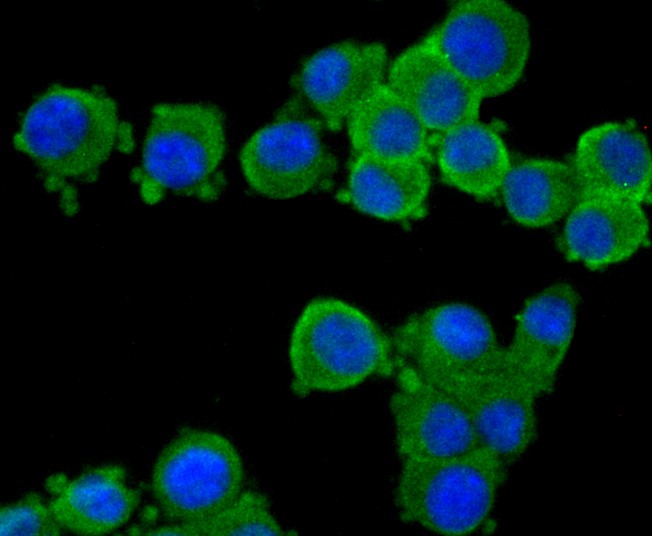
Fig3: ICC staining of CNGA4 in N2A cells (green). Formalin fixed cells were permeabilized with 0.1% Triton X-100 in TBS for 10 minutes at room temperature and blocked with 1% Blocker BSA for 15 minutes at room temperature. Cells were probed with the primary antibody (ER1901-88, 1/100) for 1 hour at room temperature, washed with PBS. Alexa Fluor®488 Goat anti-Rabbit IgG was used as the secondary antibody at 1/100 dilution. The nuclear counter stain is DAPI (blue). |
|
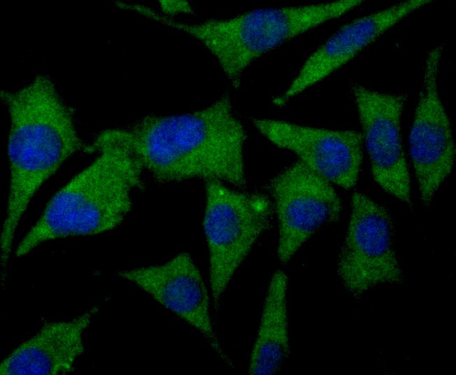
Fig4: ICC staining of CNGA4 in SHSY5Y cells (green). Formalin fixed cells were permeabilized with 0.1% Triton X-100 in TBS for 10 minutes at room temperature and blocked with 1% Blocker BSA for 15 minutes at room temperature. Cells were probed with the primary antibody (ER1901-88, 1/100) for 1 hour at room temperature, washed with PBS. Alexa Fluor®488 Goat anti-Rabbit IgG was used as the secondary antibody at 1/100 dilution. The nuclear counter stain is DAPI (blue). |
|
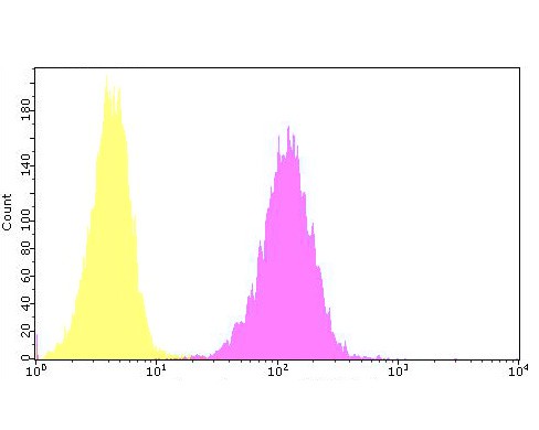
Fig5: Flow cytometric analysis of CNGA4 was done on Hela cells. The cells were fixed, permeabilized and stained with the primary antibody (ER1901-88, 1/100) (yellow). After incubation of the primary antibody at room temperature for an hour, the cells were stained with a Alexa Fluor 488-conjugated goat anti-rabbit IgG Secondary antibody at 1/500 dilution for 30 minutes.Unlabelled sample was used as a control (cells without incubation with primary antibody; purple). |
Note: All products are “FOR RESEARCH USE ONLY AND ARE NOT INTENDED FOR DIAGNOSTIC OR THERAPEUTIC USE”.