KCNMA1 Rabbit Polyclonal Antibody

cat.: ER1902-04
| Product Type: | Rabbit polyclonal IgG, primary antibodies |
|---|---|
| Species reactivity: | Human, Mouse, Rabbit |
| Applications: | WB, IF-Cell, FC |
| Clonality: | Polyclonal |
| Form: | Liquid |
| Storage condition: | Store at +4℃ after thawing. Aliquot store at -20℃. Avoid repeated freeze / thaw cycles. |
| Storage buffer: | 1*PBS (pH7.4), 0.2% BSA, 50% Glycerol. Preservative: 0.05% Sodium Azide. |
| Concentration: | 1ug/ul |
| Purification: | Immunogen affinity purified. |
| Molecular weight: | Predicted band size 138 kDa |
| Isotype: | IgG |
| Immunogen: | Synthetic peptide within rat KCNMA1 aa 200-300. |
| Positive control: | A549 cell lysates, A549, PC-3M, HCT116. |
| Subcellular location: | Cell membrane. |
| Recommended Dilutions:
WB IF-Cell FC |
1:500 1:50-1:200 1:50-1:100 |
| Uniprot #: | SwissProt: Q12791 Human | Q08460 Mouse | Q62976 Rat |
| Alternative names: | subfamily M subunit alpha-1 BK channel BKCA alpha BKCA alpha subunit BKTM Calcium-activated potassium channel Calcium-activated potassium channel subunit alpha-1 Drosophila slowpoke like hSlo K(VCA)alpha KCa1.1 KCMA1_HUMAN KCNMA KCNMA1 Maxi K channel Maxi Potassium channel alpha MaxiK SAKCA SLO alpha SLO Slo homolog Slo-alpha Slo1 Slowpoke homolog |
Images
|
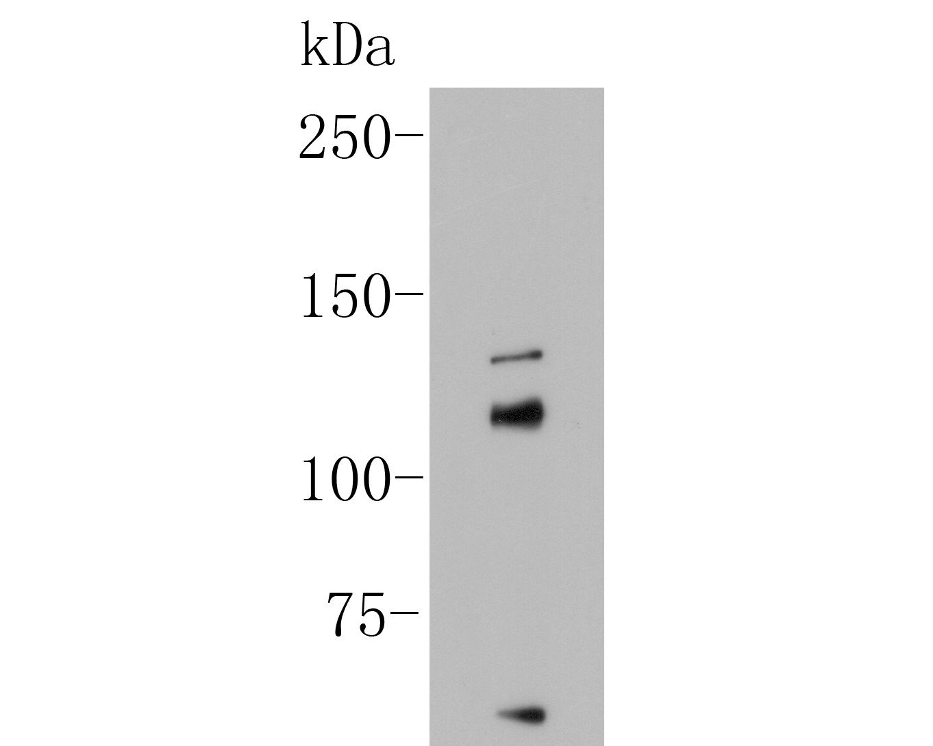
Fig1: Western blot analysis of on A549 cell lysate. Proteins were transferred to a PVDF membrane and blocked with 5% BSA in PBS for 1 hour at room temperature. The primary antibody (ER1902-04, 1/500) was used in 5% BSA at room temperature for 2 hours. Goat Anti-Rabbit IgG - HRP Secondary Antibody (HA1001) at 1:5,000 dilution was used for 1 hour at room temperature. |
|
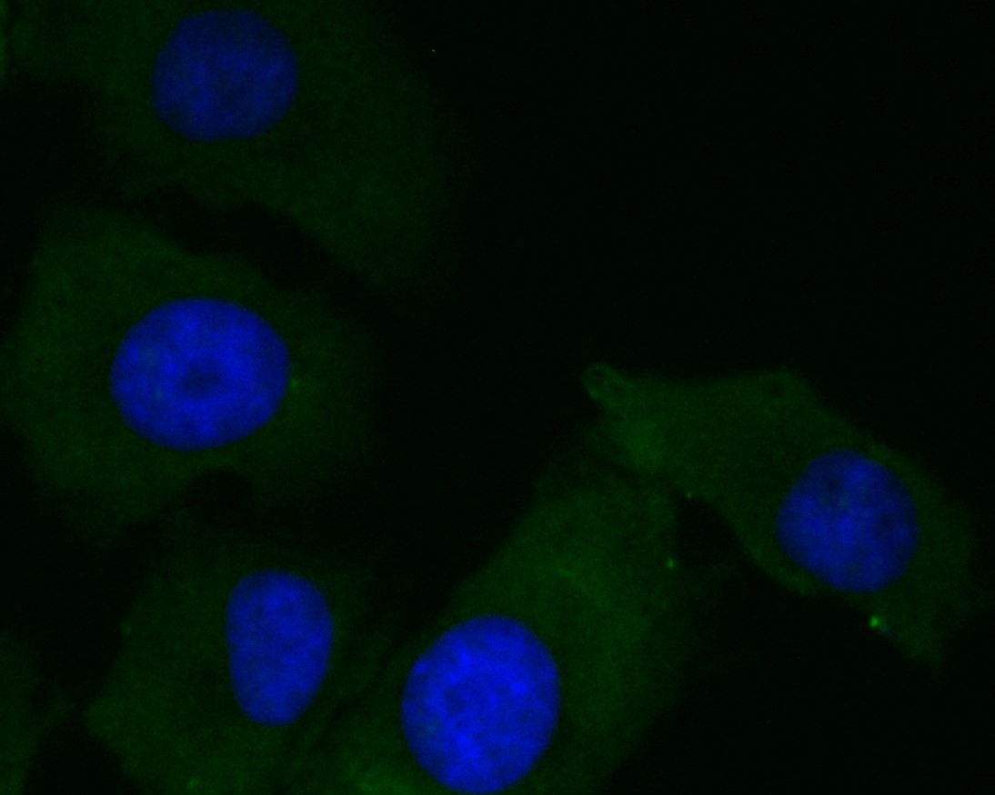
Fig2: ICC staining of KCNMA1 in A549 cells (green). Formalin fixed cells were permeabilized with 0.1% Triton X-100 in TBS for 10 minutes at room temperature and blocked with 1% Blocker BSA for 15 minutes at room temperature. Cells were probed with the primary antibody (ER1902-04, 1/100) for 1 hour at room temperature, washed with PBS. Alexa Fluor®488 Goat anti-Rabbit IgG was used as the secondary antibody at 1/100 dilution. The nuclear counter stain is DAPI (blue) |
|
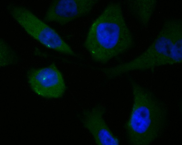
Fig3: ICC staining of KCNMA1 in PC-3M cells (green). Formalin fixed cells were permeabilized with 0.1% Triton X-100 in TBS for 10 minutes at room temperature and blocked with 1% Blocker BSA for 15 minutes at room temperature. Cells were probed with the primary antibody (ER1902-04, 1/100) for 1 hour at room temperature, washed with PBS. Alexa Fluor®488 Goat anti-Rabbit IgG was used as the secondary antibody at 1/100 dilution. The nuclear counter stain is DAPI (blue) |
|
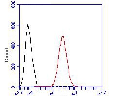
Fig4: Flow cytometric analysis of KCNMA1 was done on HCT116 cells. The cells were fixed, permeabilized and stained with the primary antibody (ER1902-04, 1/100) (red). After incubation of the primary antibody at room temperature for an hour, the cells were stained with a Alexa Fluor 488-conjugated goat anti-rabbit IgG Secondary antibody at 1/500 dilution for 30 minutes.Unlabelled sample was used as a control (cells without incubation with primary antibody; black). |
Note: All products are “FOR RESEARCH USE ONLY AND ARE NOT INTENDED FOR DIAGNOSTIC OR THERAPEUTIC USE”.