KCNMA1 Rabbit Polyclonal Antibody

cat.: ER1902-05
| Product Type: | Rabbit polyclonal IgG, primary antibodies |
|---|---|
| Species reactivity: | Human, Mouse, Rat |
| Applications: | WB, IF-Cell, IHC-P, FC |
| Clonality: | Polyclonal |
| Form: | Liquid |
| Storage condition: | Store at +4℃ after thawing. Aliquot store at -20℃. Avoid repeated freeze / thaw cycles. |
| Storage buffer: | 1*PBS (pH7.4), 0.2% BSA, 50% Glycerol. Preservative: 0.05% Sodium Azide. |
| Concentration: | 1ug/ul |
| Purification: | Immunogen affinity purified. |
| Molecular weight: | Predicted band size 138 kDa |
| Isotype: | IgG |
| Immunogen: | Synthetic peptide within rat KCNMA1 aa 100-300. |
| Positive control: | Rat brain lysates, A549, rat cerebellum tissue, human kidney tissue, human small intestine tissue, mouse colon tissue, HCT116. |
| Subcellular location: | Cell membrane, Endoplasmic reticulum, Membrane. |
| Recommended Dilutions:
WB IF-Cell IHC-P FC |
1:500-1:1000 1:50-1:200 1:50-1:200 1:50-1:100 |
| Uniprot #: | SwissProt: Q12791 Human | Q08460 Mouse | Q62976 Rat |
| Alternative names: | subfamily M subunit alpha-1 BK channel BKCA alpha BKCA alpha subunit BKTM Calcium-activated potassium channel Calcium-activated potassium channel subunit alpha-1 Drosophila slowpoke like hSlo K(VCA)alpha KCa1.1 KCMA1_HUMAN KCNMA KCNMA1 Maxi K channel Maxi Potassium channel alpha MaxiK SAKCA SLO alpha SLO Slo homolog Slo-alpha Slo1 Slowpoke homolog |
Images
|
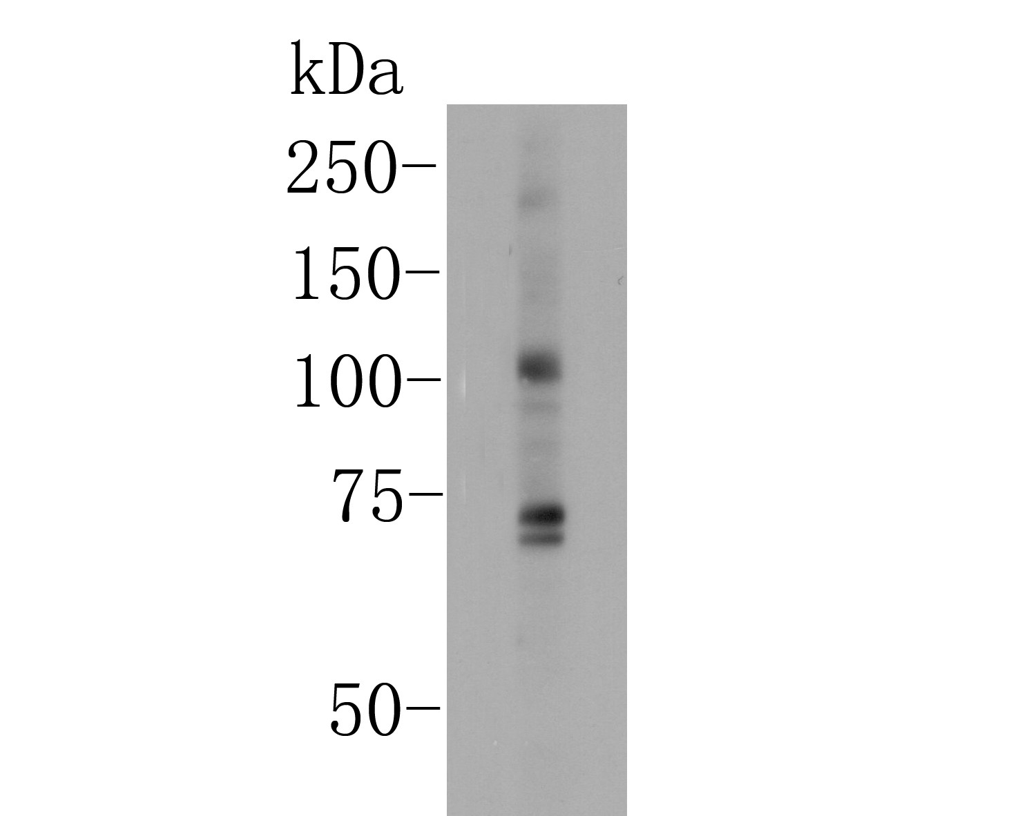
Fig1: Western blot analysis of KCNMA1 on rat brain lysates. Proteins were transferred to a PVDF membrane and blocked with 5% BSA in PBS for 1 hour at room temperature. The primary antibody (ER1902-05, 1/500) was used in 5% BSA at room temperature for 2 hours. Goat Anti-Rabbit IgG - HRP Secondary Antibody (HA1001) at 1:5,000 dilution was used for 1 hour at room temperature. |
|
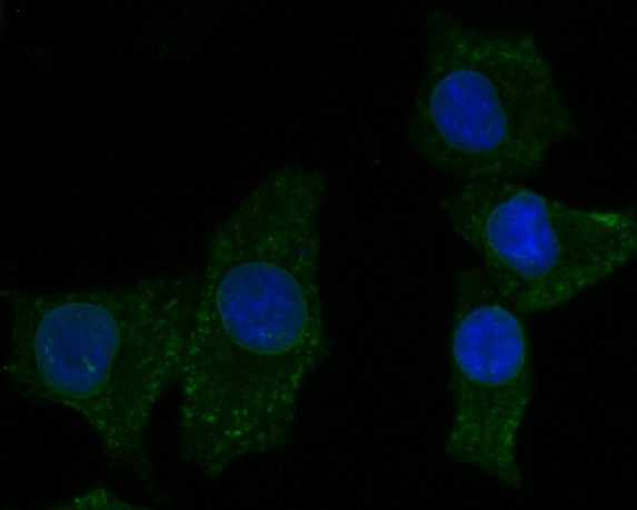
Fig2: ICC staining of KCNMA1 in A549 cells (green). Formalin fixed cells were permeabilized with 0.1% Triton X-100 in TBS for 10 minutes at room temperature and blocked with 1% Blocker BSA for 15 minutes at room temperature. Cells were probed with the primary antibody (ER1902-05, 1/50) for 1 hour at room temperature, washed with PBS. Alexa Fluor®488 Goat anti-Rabbit IgG was used as the secondary antibody at 1/1,000 dilution. The nuclear counter stain is DAPI (blue). |
|
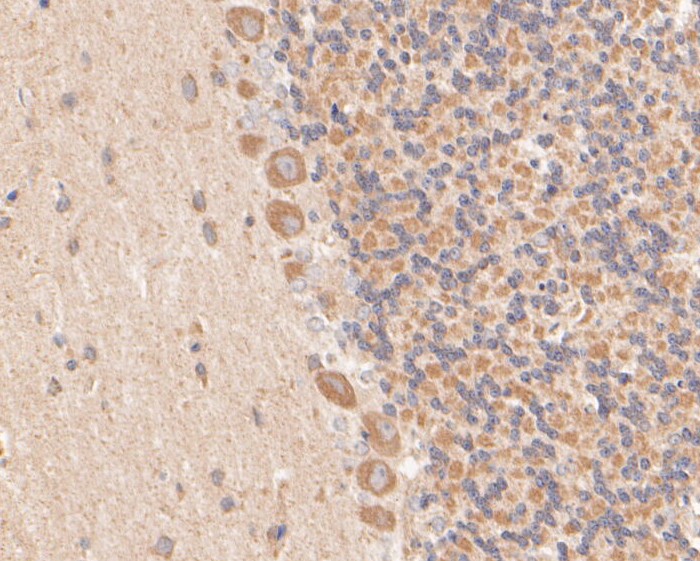
Fig3: Immunohistochemical analysis of paraffin-embedded rat cerebellum tissue using anti-KCNMA1 antibody. The section was pre-treated using heat mediated antigen retrieval with Tris-EDTA buffer (pH 8.0-8.4) for 20 minutes.The tissues were blocked in 5% BSA for 30 minutes at room temperature, washed with ddH2O and PBS, and then probed with the primary antibody (ER1902-05, 1/50) for 30 minutes at room temperature. The detection was performed using an HRP conjugated compact polymer system. DAB was used as the chromogen. Tissues were counterstained with hematoxylin and mounted with DPX. |

|
Fig4: Immunohistochemical analysis of paraffin-embedded human kidney tissue using anti-KCNMA1 antibody. The section was pre-treated using heat mediated antigen retrieval with Tris-EDTA buffer (pH 8.0-8.4) for 20 minutes.The tissues were blocked in 5% BSA for 30 minutes at room temperature, washed with ddH2O and PBS, and then probed with the primary antibody (ER1902-05, 1/50) for 30 minutes at room temperature. The detection was performed using an HRP conjugated compact polymer system. DAB was used as the chromogen. Tissues were counterstained with hematoxylin and mounted with DPX. |
|
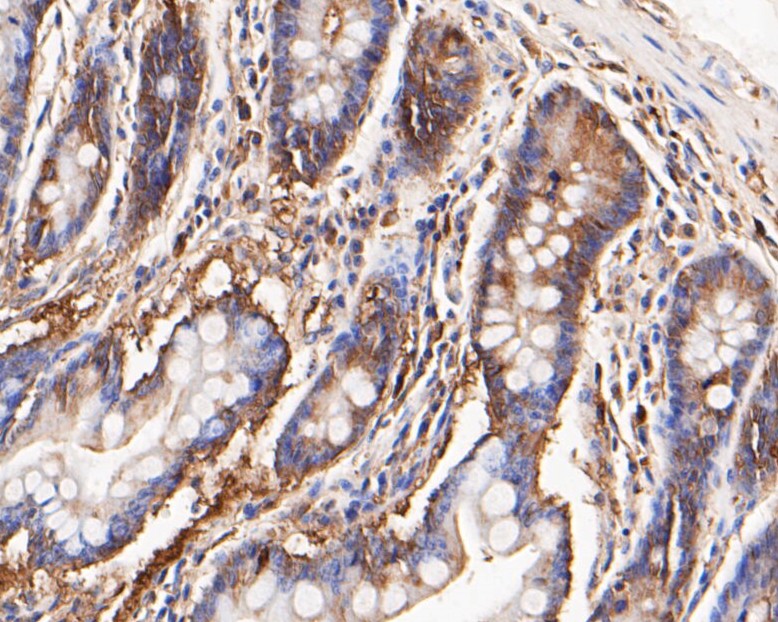
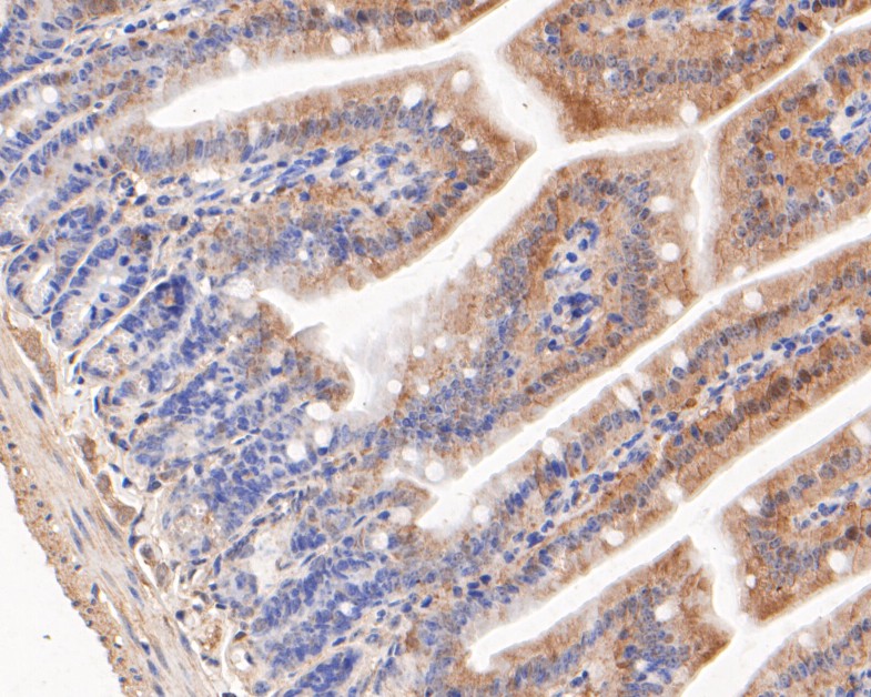
Fig5: Immunohistochemical analysis of paraffin-embedded human small intestine tissue using anti-KCNMA1 antibody. The section was pre-treated using heat mediated antigen retrieval with Tris-EDTA buffer (pH 8.0-8.4) for 20 minutes.The tissues were blocked in 5% BSA for 30 minutes at room temperature, washed with ddH2O and PBS, and then probed with the primary antibody (ER1902-05, 1/50) for 30 minutes at room temperature. The detection was performed using an HRP conjugated compact polymer system. DAB was used as the chromogen. Tissues were counterstained with hematoxylin and mounted with DPX. |
|
Fig6: Immunohistochemical analysis of paraffin-embedded mouse colon tissue using anti-KCNMA1 antibody. The section was pre-treated using heat mediated antigen retrieval with Tris-EDTA buffer (pH 8.0-8.4) for 20 minutes.The tissues were blocked in 5% BSA for 30 minutes at room temperature, washed with ddH2O and PBS, and then probed with the primary antibody (ER1902-05, 1/50) for 30 minutes at room temperature. The detection was performed using an HRP conjugated compact polymer system. DAB was used as the chromogen. Tissues were counterstained with hematoxylin and mounted with DPX. |
|
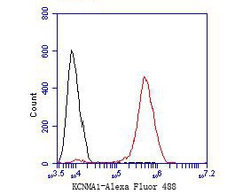
Fig7: Flow cytometric analysis of KCNMA1 was done on HCT116 cells. The cells were fixed, permeabilized and stained with the primary antibody (ER1902-05, 1/50) (red). After incubation of the primary antibody at room temperature for an hour, the cells were stained with a Alexa Fluor 488-conjugated Goat anti-Rabbit IgG Secondary antibody at 1/1000 dilution for 30 minutes.Unlabelled sample was used as a control (cells without incubation with primary antibody; black). |
Note: All products are “FOR RESEARCH USE ONLY AND ARE NOT INTENDED FOR DIAGNOSTIC OR THERAPEUTIC USE”.