TMX4 Rabbit Polyclonal Antibody

cat.: ER1902-31
| Product Type: | Rabbit polyclonal IgG, primary antibodies |
|---|---|
| Species reactivity: | Human |
| Applications: | WB, IHC-P, FC |
| Clonality: | Polyclonal |
| Form: | Liquid |
| Storage condition: | Shipped at 4℃. Store at +4℃ short term (1-2 weeks). It is recommended to aliquot into single-use upon delivery. Store at -20℃ long term. |
| Storage buffer: | 1*PBS (pH7.4), 0.2% BSA, 50% Glycerol. Preservative: 0.05% Sodium Azide. |
| Concentration: | 1ug/ul |
| Purification: | Immunogen affinity purified. |
| Molecular weight: | Predicted band size: 39 kDa |
| Isotype: | IgG |
| Immunogen: | Synthetic peptide within Human TMX4 aa 300-349 / 349. |
| Positive control: | Daudi cell lysate, human skin tissue lysate, human tonsil tissue, human liver carcinoma tissue, human colon carcinoma tissue, human breast carcinoma tissue, SH-SY5Y. |
| Subcellular location: | Nucleus inner membrane. |
| Recommended Dilutions:
WB IHC-P FC |
1:500-1:1,000 1:50-1:200 1:50-1:100 |
| Uniprot #: | SwissProt: Q9H1E5 Human |
| Alternative names: | DJ971N18.2 KIAA1162 PDIA14 Protein disulfide isomerase family A 14 PSEC0095 Thioredoxin domain containing 13 Thioredoxin domain containing protein 13 Thioredoxin related transmembrane protein 4 TMX4 TXNDC 13 UNQ475/PRO938 |
Images
|
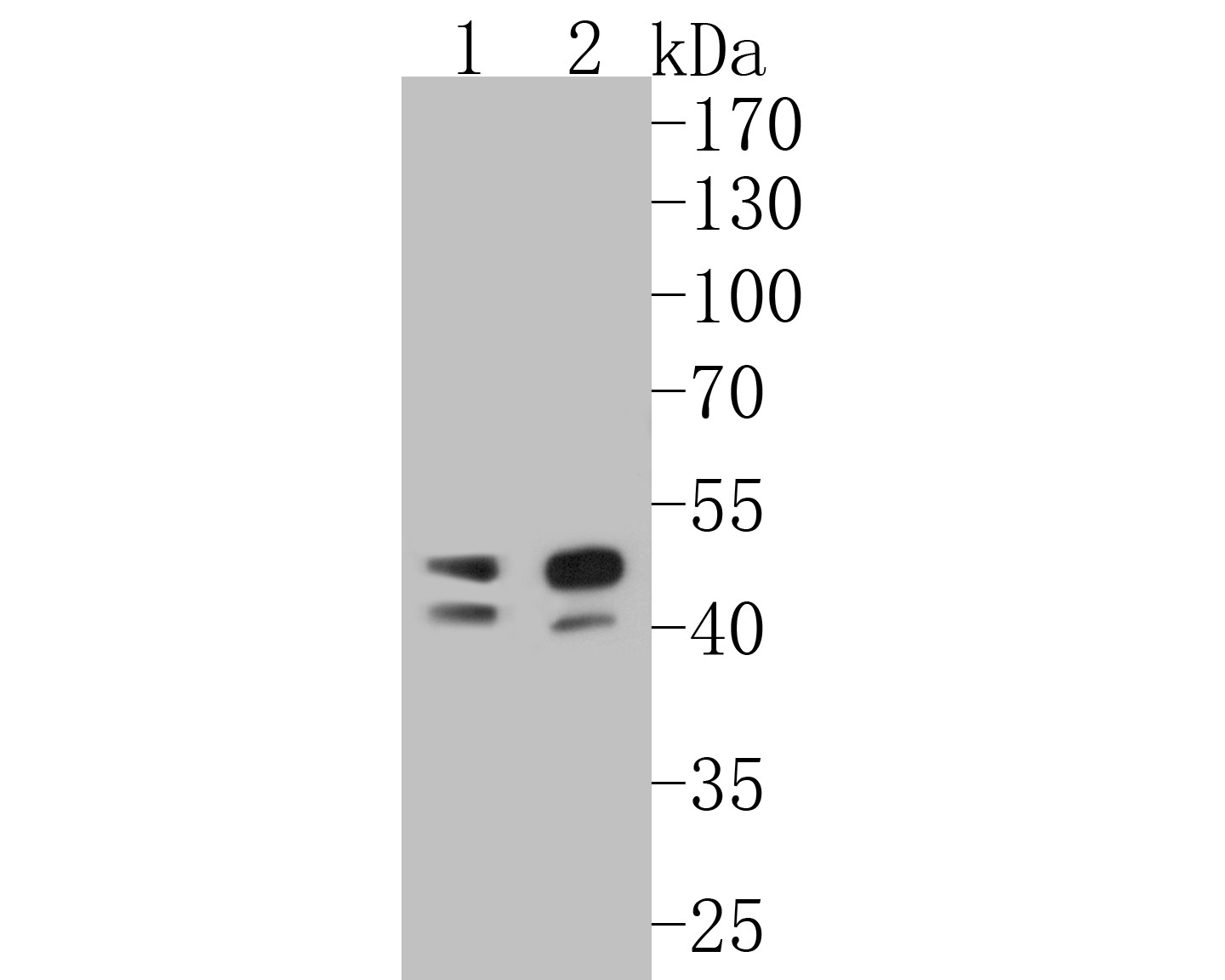
Fig1:
Western blot analysis of TMX4 on different lysates. Proteins were transferred to a PVDF membrane and blocked with 5% BSA in PBS for 1 hour at room temperature. The primary antibody (ER1902-31, 1/500) was used in 5% BSA at room temperature for 2 hours. Goat Anti-Rabbit IgG - HRP Secondary Antibody (HA1001) at 1:5,000 dilution was used for 1 hour at room temperature. Positive control: Lane 1: Daudi cell lysate Lane 2: Human skin tissue lysate |
|
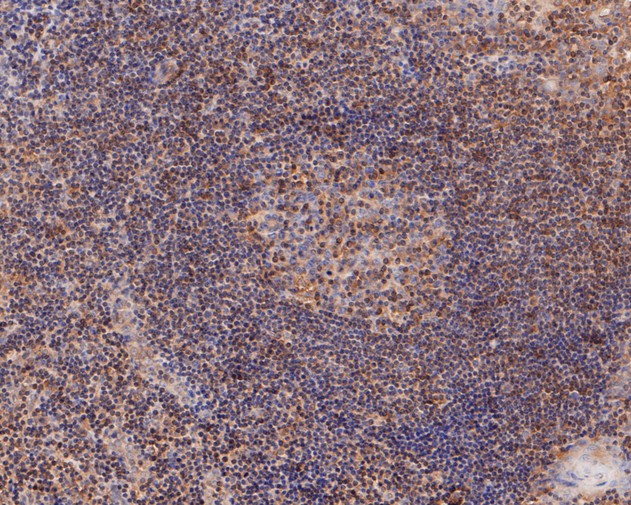
Fig2: Immunohistochemical analysis of paraffin-embedded human tonsil tissue using anti-TMX4 antibody. The section was pre-treated using heat mediated antigen retrieval with Tris-EDTA buffer (pH 8.0-8.4) for 20 minutes.The tissues were blocked in 5% BSA for 30 minutes at room temperature, washed with ddH2O and PBS, and then probed with the primary antibody (ER1902-31, 1/50) for 30 minutes at room temperature. The detection was performed using an HRP conjugated compact polymer system. DAB was used as the chromogen. Tissues were counterstained with hematoxylin and mounted with DPX. |
|
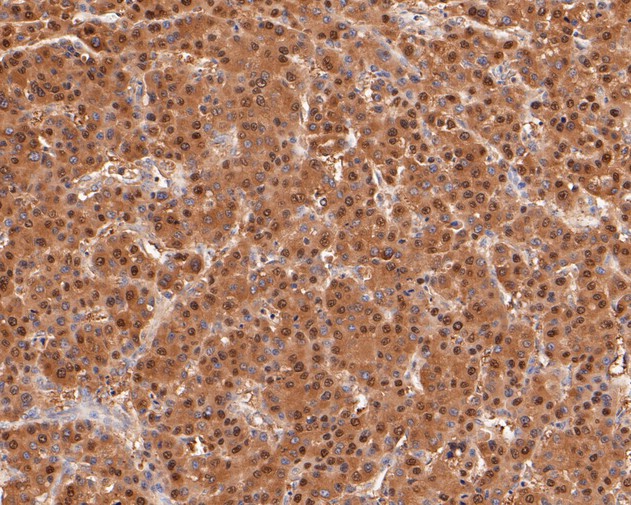
Fig3: Immunohistochemical analysis of paraffin-embedded human liver carcinoma tissue using anti-TMX4 antibody. The section was pre-treated using heat mediated antigen retrieval with Tris-EDTA buffer (pH 8.0-8.4) for 20 minutes.The tissues were blocked in 5% BSA for 30 minutes at room temperature, washed with ddH2O and PBS, and then probed with the primary antibody (ER1902-31, 1/50) for 30 minutes at room temperature. The detection was performed using an HRP conjugated compact polymer system. DAB was used as the chromogen. Tissues were counterstained with hematoxylin and mounted with DPX. |
|
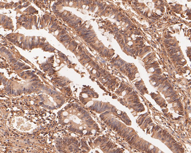
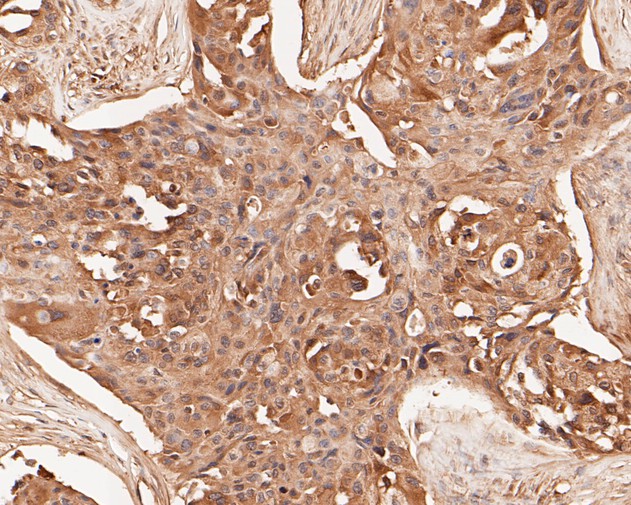
Fig4: Immunohistochemical analysis of paraffin-embedded human colon carcinoma tissue using anti-TMX4 antibody. The section was pre-treated using heat mediated antigen retrieval with Tris-EDTA buffer (pH 8.0-8.4) for 20 minutes.The tissues were blocked in 5% BSA for 30 minutes at room temperature, washed with ddH2O and PBS, and then probed with the primary antibody (ER1902-31, 1/50) for 30 minutes at room temperature. The detection was performed using an HRP conjugated compact polymer system. DAB was used as the chromogen. Tissues were counterstained with hematoxylin and mounted with DPX. |
|
Fig5: Immunohistochemical analysis of paraffin-embedded human breast carcinoma tissue using anti-TMX4 antibody. The section was pre-treated using heat mediated antigen retrieval with Tris-EDTA buffer (pH 8.0-8.4) for 20 minutes.The tissues were blocked in 5% BSA for 30 minutes at room temperature, washed with ddH2O and PBS, and then probed with the primary antibody (ER1902-31, 1/50) for 30 minutes at room temperature. The detection was performed using an HRP conjugated compact polymer system. DAB was used as the chromogen. Tissues were counterstained with hematoxylin and mounted with DPX. |

|
Fig6: Flow cytometric analysis of TMX4 was done on SH-SY5Y cells. The cells were fixed, permeabilized and stained with the primary antibody (ER1902-31, 1/50) (red). After incubation of the primary antibody at room temperature for an hour, the cells were stained with a Alexa Fluor 488-conjugated Goat anti-Rabbit IgG Secondary antibody at 1/1,000 dilution for 30 minutes.Unlabelled sample was used as a control (cells without incubation with primary antibody; black). |
Note: All products are “FOR RESEARCH USE ONLY AND ARE NOT INTENDED FOR DIAGNOSTIC OR THERAPEUTIC USE”.