IL-17A Rabbit Polyclonal Antibody

cat.: ER1902-37
| Product Type: | Rabbit polyclonal IgG, primary antibodies |
|---|---|
| Species reactivity: | Human, Mouse, Rat |
| Applications: | WB, IHC-P, FC, mIHC |
| Clonality: | Polyclonal |
| Form: | Liquid |
| Storage condition: | Shipped at 4℃. Store at +4℃ short term (1-2 weeks). It is recommended to aliquot into single-use upon delivery. Store at -20℃ long term. |
| Storage buffer: | 1*PBS (pH7.4), 0.2% BSA, 50% Glycerol. Preservative: 0.05% Sodium Azide. |
| Concentration: | 1ug/ul |
| Purification: | Immunogen affinity purified. |
| Molecular weight: | Predicted band size: 18 kDa |
| Isotype: | IgG |
| Immunogen: | Recombinant protein within Human IL17 aa 1-155 / 155. |
| Positive control: | Recombinant protein lysates, Rat stomach tissue lysates, Human skeletal muscle tissue lysate, human fetal skeletal muscle tissue, human esophagus tissue, mouse colon tissue, Jurkat, mouse spleen tissue. |
| Subcellular location: | Secreted. |
| Recommended Dilutions:
WB IHC-P FC mIHC |
1:500 1:50-1:800 1:50-1:100 1:2,000 |
| Uniprot #: | SwissProt: Q16552 Human | Q62386 Mouse |
| Alternative names: | CTLA 8 CTLA-8 CTLA8 Cytotoxic T lymphocyte associated antigen 8 Cytotoxic T lymphocyte associated protein 8 Cytotoxic T lymphocyte associated serine esterase 8 Cytotoxic T-lymphocyte-associated antigen 8 IL 17A IL-17 IL-17A IL17 IL17_HUMAN Il17a Interleukin 17 (cytotoxic T lymphocyte associated serine esterase 8) Interleukin 17A Interleukin-17A Interleukin17 Interleukin17A OTTHUMP00000016597 OTTMUSP00000046003 |
Images
|
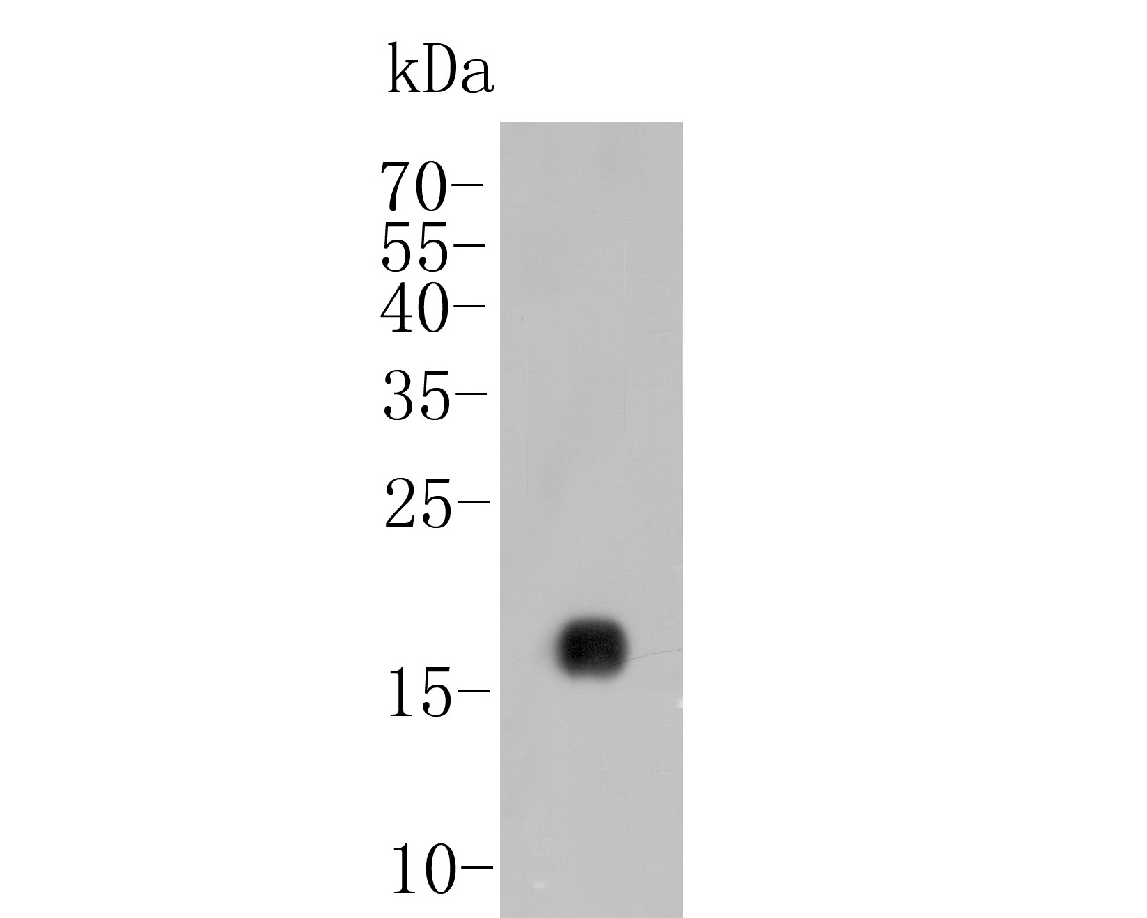
Fig1: Western blot analysis of IL-17A on recombinant protein lysate. Proteins were transferred to a PVDF membrane and blocked with 5% BSA in PBS for 1 hour at room temperature. The primary antibody (ER1902-37, 1/20,000) was used in 5% BSA at room temperature for 2 hours. Goat Anti-Rabbit IgG - HRP Secondary Antibody (HA1001) at 1:5,000 dilution was used for 1 hour at room temperature. |
|
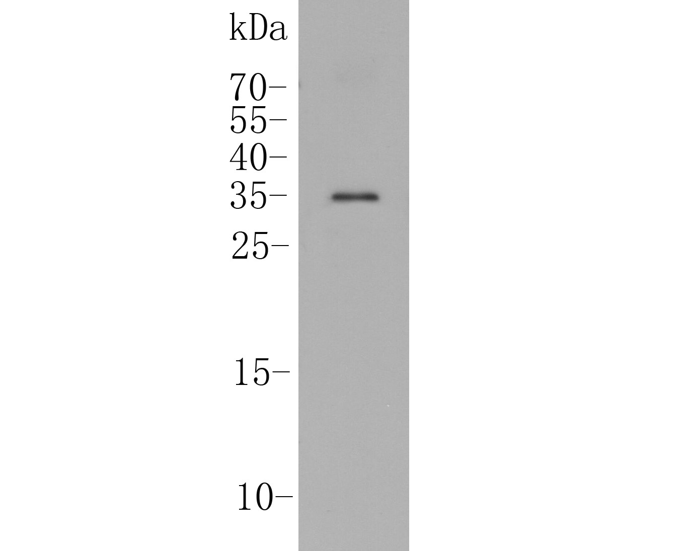
Fig2: Western blot analysis of IL-17A on Rat stomach tissue lysate. Proteins were transferred to a PVDF membrane and blocked with 5% BSA in PBS for 1 hour at room temperature. The primary antibody (ER1902-37, 1/500) was used in 5% BSA at room temperature for 2 hours. Goat Anti-Rabbit IgG - HRP Secondary Antibody (HA1001) at 1:5,000 dilution was used for 1 hour at room temperature. |
|
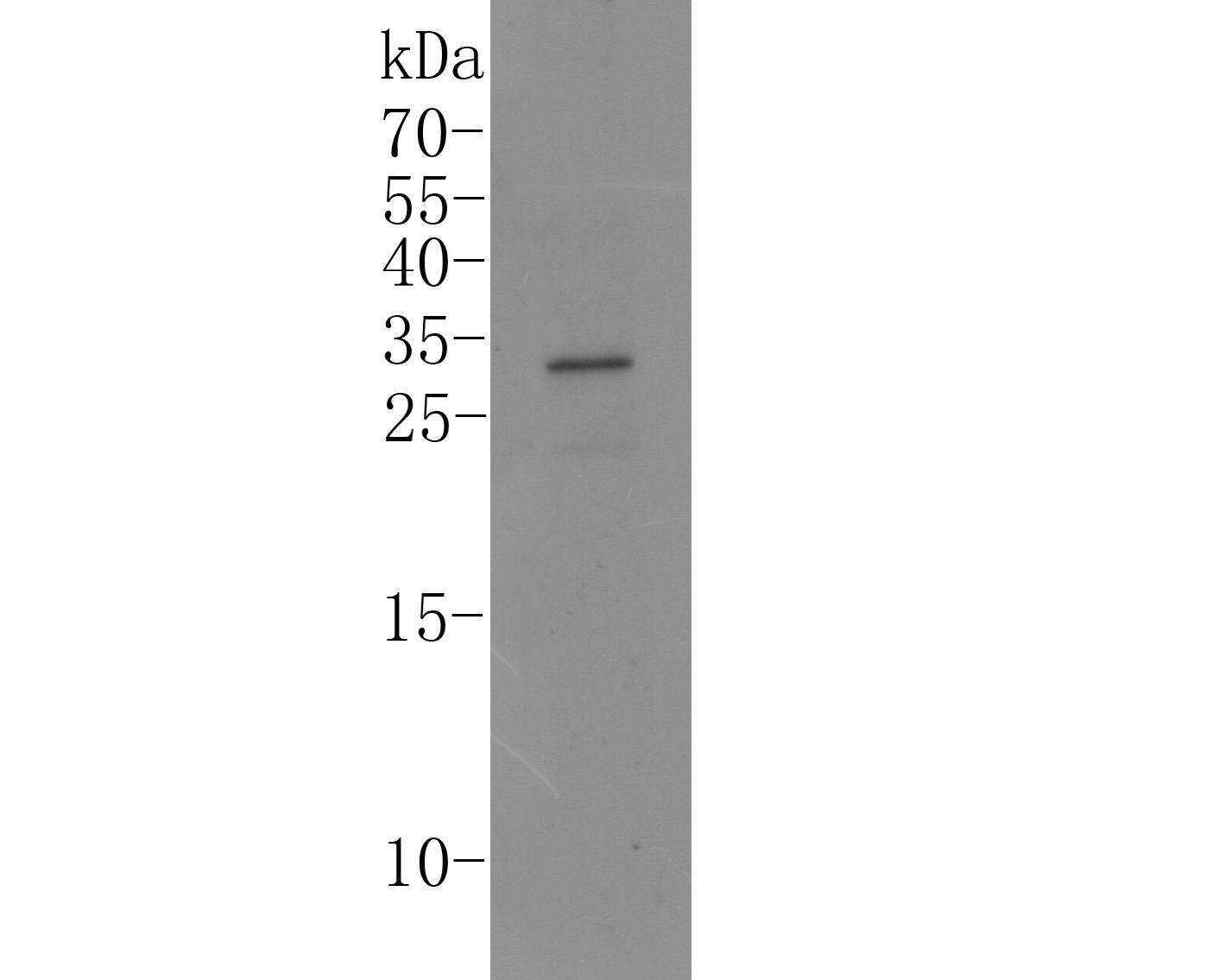
Fig3: Western blot analysis of IL-17A on Human skeletal muscle tissue lysate. Proteins were transferred to a PVDF membrane and blocked with 5% BSA in PBS for 1 hour at room temperature. The primary antibody (ER1902-37, 1/500) was used in 5% BSA at room temperature for 2 hours. Goat Anti-Rabbit IgG - HRP Secondary Antibody (HA1001) at 1:5,000 dilution was used for 1 hour at room temperature. |
|
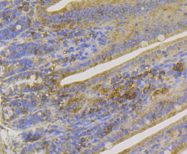
Fig4: Immunohistochemical analysis of paraffin-embedded mouse colon tissue using anti-IL-17A antibody. The section was pre-treated using heat mediated antigen retrieval with Tris-EDTA buffer (pH 8.0-8.4) for 20 minutes.The tissues were blocked in 5% BSA for 30 minutes at room temperature, washed with ddH2O and PBS, and then probed with the primary antibody (ER1902-37, 1/100) for 30 minutes at room temperature. The detection was performed using an HRP conjugated compact polymer system. DAB was used as the chromogen. Tissues were counterstained with hematoxylin and mounted with DPX. |
|
Fig5:
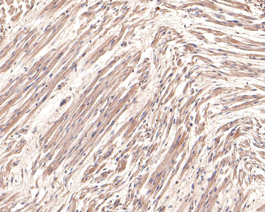
Immunohistochemical analysis of paraffin-embedded human fetal skeletal muscle tissue with Rabbit anti-IL-17A antibody (ER1902-37) at 1/800 dilution. The section was pre-treated using heat mediated antigen retrieval with Tris-EDTA buffer (pH 9.0) for 20 minutes. The tissues were blocked in 1% BSA for 20 minutes at room temperature, washed with ddH2O and PBS, and then probed with the primary antibody (ER1902-37) at 1/800 dilution for 1 hour at room temperature. The detection was performed using an HRP conjugated compact polymer system. DAB was used as the chromogen. Tissues were counterstained with hematoxylin and mounted with DPX. |
|
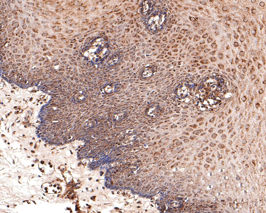
Fig6:
Immunohistochemical analysis of paraffin-embedded human esophagus tissue with Rabbit anti-IL-17A antibody (ER1902-37) at 1/800 dilution. The section was pre-treated using heat mediated antigen retrieval with Tris-EDTA buffer (pH 9.0) for 20 minutes. The tissues were blocked in 1% BSA for 20 minutes at room temperature, washed with ddH2O and PBS, and then probed with the primary antibody (ER1902-37) at 1/800 dilution for 1 hour at room temperature. The detection was performed using an HRP conjugated compact polymer system. DAB was used as the chromogen. Tissues were counterstained with hematoxylin and mounted with DPX. |
|
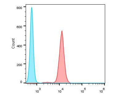
Fig7: Flow cytometric analysis of IL-17A was done on Jurkat cells. The cells were fixed, permeabilized and stained with the primary antibody (ER1902-37, 1/100) (red). After incubation of the primary antibody at room temperature for an hour, the cells were stained with a Alexa Fluor 488-conjugated goat anti-rabbit IgG Secondary antibody at 1/500 dilution for 30 minutes.Unlabelled sample was used as a control (cells without incubation with primary antibody; blue). |
|
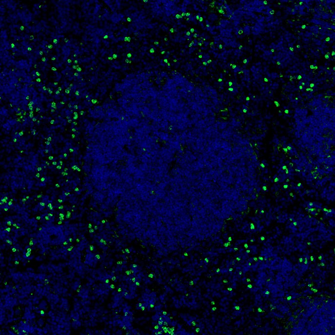
Fig8: mIHC analysis of mouse spleen tissue (Formalin/PFA-fixed paraffin-embedded sections) with Rabbit anti-IL-17A antibody (ER1902-37) at 1/2,000 dilution. The immunostaining was performed with the IRISKit® HyperView mTSA Kit (MH900206). Heat mediated antigen retrieval with Tris-EDTA buffer (pH 9.0) for 30 mins at 95℃. DAPI (blue) was used as a nuclear counter stain. Image acquisition was performed with Olympus VS200 Slide Scanner. |
Note: All products are “FOR RESEARCH USE ONLY AND ARE NOT INTENDED FOR DIAGNOSTIC OR THERAPEUTIC USE”.