SynGAP Rabbit Polyclonal Antibody

cat.: ER1902-57
| Product Type: | Rabbit polyclonal IgG, primary antibodies |
|---|---|
| Species reactivity: | Human, Mouse, Rat |
| Applications: | WB, IHC-P, FC |
| Clonality: | Polyclonal |
| Form: | Liquid |
| Storage condition: | Shipped at 4℃. Store at +4℃ short term (1-2 weeks). It is recommended to aliquot into single-use upon delivery. Store at -20℃ long term. |
| Storage buffer: | 1*PBS (pH7.4), 0.2% BSA, 50% Glycerol. Preservative: 0.05% Sodium Azide. |
| Concentration: | 1ug/ul |
| Purification: | Immunogen affinity purified. |
| Molecular weight: | 148 kDa |
| Isotype: | IgG |
| Immunogen: | Recombinant protein within human SynGAP aa 1100-1250. |
| Positive control: | K562 cell lysate, mouse brain tissue lysate, human terus tissue, mouse brain tissue, SW620. |
| Subcellular location: | Cytosol. |
| Recommended Dilutions:
WB IHC-P FC |
1:500-1:2,000 1:50-1:100 1:50-1:100 |
| Uniprot #: | SwissProt: Q96PV0 Human | Q9QUH6 Rat | F6SEU4 Mouse |
| Alternative names: | DKFZp761G1421 KIAA1938 MRD5 Neuronal RasGAP OTTHUMP00000064825 p135 SynGAP Ras GTPase activating protein SynGAP Ras GTPase-activating protein SynGAP RASA 1 RASA 5 RASA1 RASA5 SYGP1_HUMAN Synaptic Ras GAP 1 Synaptic Ras GTPase activating protein 1 Synaptic Ras GTPase activating protein 1 homolog Synaptic Ras GTPase activating protein 135kDa Synaptic Ras GTPase activating protein Synaptic Ras GTPase-activating protein 1 Synaptic Ras-GAP 1 SYNGAP 1 SYNGAP1 |
Images
|
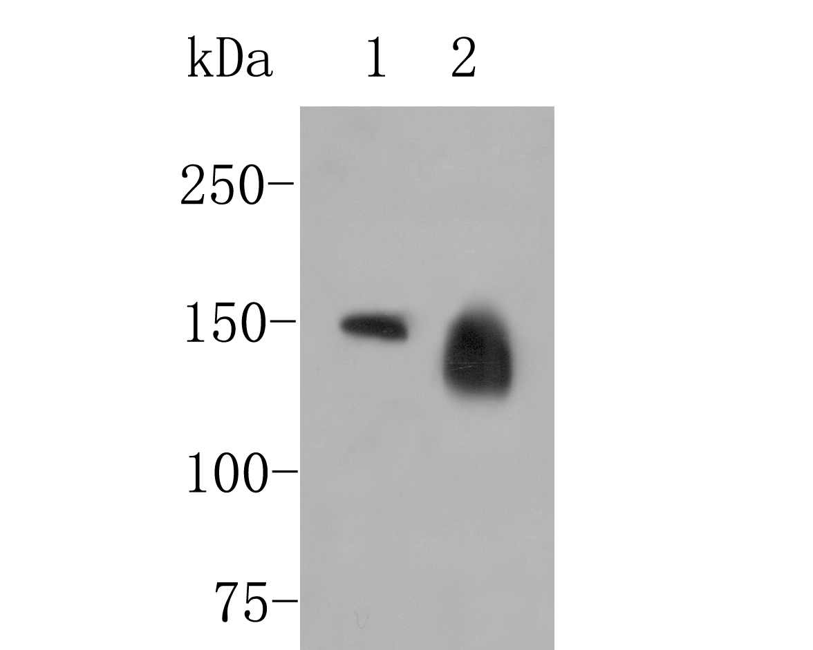
Fig1:
Western blot analysis of SynGAP on different lysates. Proteins were transferred to a PVDF membrane and blocked with 5% BSA in PBS for 1 hour at room temperature. The primary antibody (ER1902-57, 1/500) was used in 5% BSA at room temperature for 2 hours. Goat Anti-Rabbit IgG - HRP Secondary Antibody (HA1001) at 1:5,000 dilution was used for 1 hour at room temperature. Positive control: Lane 1: K562 cell lysate Lane 2: Mouse brain tissue lysate |
|
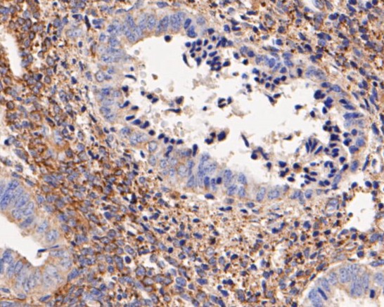
Fig2: Immunohistochemical analysis of paraffin-embedded human terus tissue using anti-SynGAP antibody. The section was pre-treated using heat mediated antigen retrieval with Tris-EDTA buffer (pH 8.0-8.4) for 20 minutes.The tissues were blocked in 5% BSA for 30 minutes at room temperature, washed with ddH2O and PBS, and then probed with the primary antibody (ER1902-57, 1/100) for 30 minutes at room temperature. The detection was performed using an HRP conjugated compact polymer system. DAB was used as the chromogen. Tissues were counterstained with hematoxylin and mounted with DPX. |
|
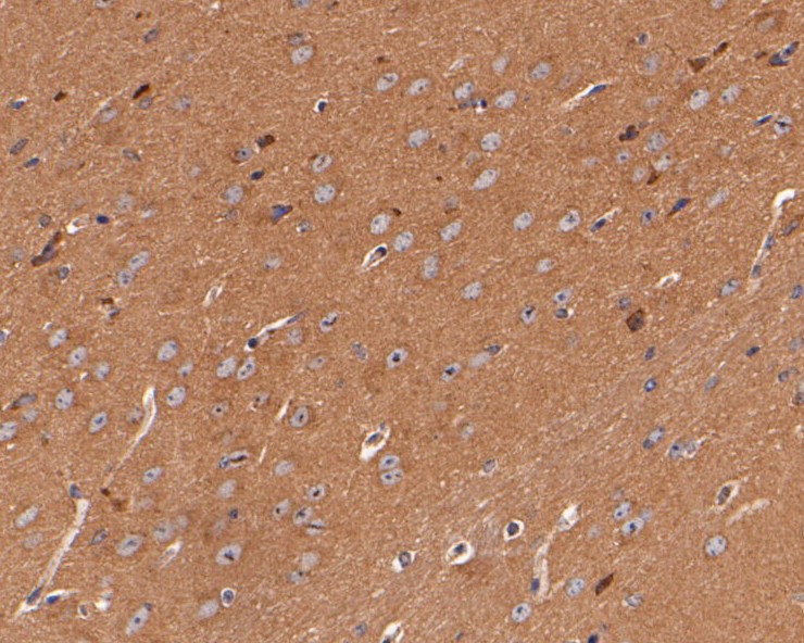
Fig3: Immunohistochemical analysis of paraffin-embedded mouse brain tissue using anti-SynGAP antibody. The section was pre-treated using heat mediated antigen retrieval with Tris-EDTA buffer (pH 8.0-8.4) for 20 minutes.The tissues were blocked in 5% BSA for 30 minutes at room temperature, washed with ddH2O and PBS, and then probed with the primary antibody (ER1902-57, 1/100) for 30 minutes at room temperature. The detection was performed using an HRP conjugated compact polymer system. DAB was used as the chromogen. Tissues were counterstained with hematoxylin and mounted with DPX. |
|
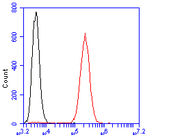
Fig4: Flow cytometric analysis of SynGAP was done on SW620 cells. The cells were fixed, permeabilized and stained with the primary antibody (ER1902-57, 1/100) (red). After incubation of the primary antibody at room temperature for an hour, the cells were stained with a Alexa Fluor 488-conjugated goat anti-rabbit IgG Secondary antibody at 1/500 dilution for 30 minutes.Unlabelled sample was used as a control (cells without incubation with primary antibody; black). |
Note: All products are “FOR RESEARCH USE ONLY AND ARE NOT INTENDED FOR DIAGNOSTIC OR THERAPEUTIC USE”.