CDK5RAP3 Rabbit Polyclonal Antibody

cat.: ER1902-72
| Product Type: | Rabbit polyclonal IgG, primary antibodies |
|---|---|
| Species reactivity: | Human, Mouse, Rat |
| Applications: | WB, IF-Cell, IHC-P |
| Clonality: | Polyclonal |
| Form: | Liquid |
| Storage condition: | Shipped at 4℃. Store at +4℃ short term (1-2 weeks). It is recommended to aliquot into single-use upon delivery. Store at -20℃ long term. |
| Storage buffer: | 1*PBS (pH7.4), 0.2% BSA, 50% Glycerol. Preservative: 0.05% Sodium Azide. |
| Concentration: | 1ug/ul |
| Purification: | Immunogen affinity purified. |
| Molecular weight: | Predicted band size: 57 kDa |
| Isotype: | IgG |
| Immunogen: | Recombinant protein within Human CDK5RAP3 aa 296-506 / 506. |
| Positive control: | HeLa cell lysate, A431 cell lysate, MCF7 cell lysate, EL4 cell lysate, PC-12 cell lysate, EL4, PC-12, mouse stomach tissue, rat stomach tissue. |
| Subcellular location: | Cytoplasm, Cytoskeleton, Nucleus. |
| Recommended Dilutions:
WB IF-Cell IHC-P |
1:5,000 1:100 1:200 |
| Uniprot #: | SwissProt: Q96JB5 Human | Q99LM2 Mouse | Q9JLH7 Rat |
| Alternative names: | C53 CDK5 activator binding protein C53 CDK5 activator-binding protein C53 CDK5 regulatory subunit associated protein 3 CDK5 regulatory subunit-associated protein 3 Cdk5rap3 CK5P3_HUMAN HSF 27 HSF27 IC53 Ischemic heart CDK5 activator binding protein C53 LXXLL/leucine zipper containing ARFbinding protein LZAP MST016 OK/SW cl.114 PP1553 Protein HSF-27 |
Images
|
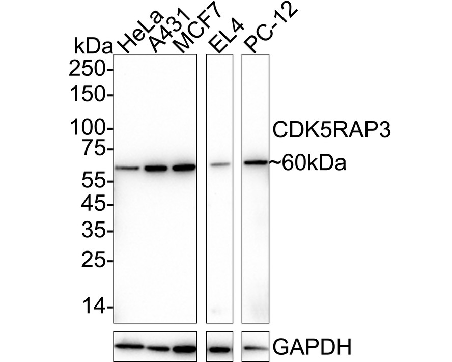
Fig1:
Western blot analysis of CDK5RAP3 on different lysates with Rabbit anti-CDK5RAP3 antibody (ER1902-72) at 1/5,000 dilution. Lane 1: HeLa cell lysate Lane 2: A431 cell lysate Lane 3: MCF7 cell lysate Lane 4: EL4 cell lysate Lane 5: PC-12 cell lysate Lysates/proteins at 20 µg/Lane. Predicted band size: 57 kDa Observed band size: 60 kDa Exposure time: 1 minute 39 seconds; ECL: K1801; 4-20% SDS-PAGE gel. Proteins were transferred to a PVDF membrane and blocked with 5% NFDM/TBST for 1 hour at room temperature. The primary antibody (ER1902-72) at 1/5,000 dilution was used in primary antibody dilution (K1803) at 4℃ overnight. Goat Anti-Rabbit IgG - HRP Secondary Antibody (HA1001) at 1/50,000 dilution was used for 1 hour at room temperature. |
|
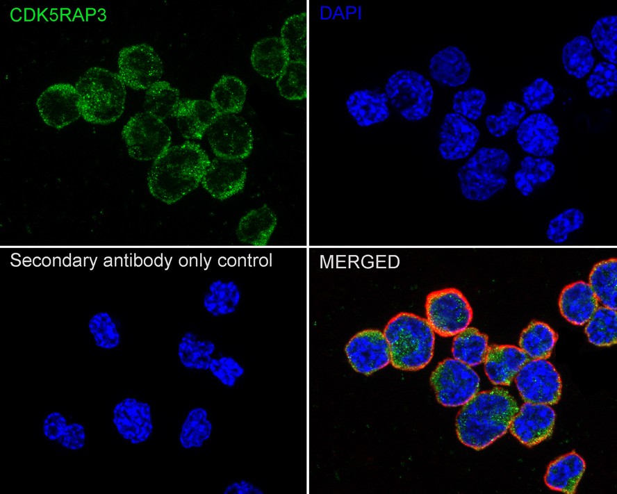
Fig2:
Immunocytochemistry analysis of EL4 cells labeling CDK5RAP3 with Rabbit anti-CDK5RAP3 antibody (ER1902-72) at 1/100 dilution. Cells were fixed in 4% paraformaldehyde for 15 minutes at room temperature, permeabilized with 0.1% Triton X-100 in PBS for 15 minutes at room temperature, then blocked with 1% BSA in 10% negative goat serum for 1 hour at room temperature. Cells were then incubated with Rabbit anti-CDK5RAP3 antibody (ER1902-72) at 1/100 dilution in 1% BSA in PBST overnight at 4 ℃. Goat Anti-Rabbit IgG H&L (iFluor™ 488, HA1121) was used as the secondary antibody at 1/1,000 dilution. PBS instead of the primary antibody was used as the secondary antibody only control. Nuclear DNA was labelled in blue with DAPI. Beta tubulin (HA601187, red) was stained at 1/100 dilution overnight at +4℃. Goat Anti-Mouse IgG H&L (iFluor™ 594, HA1126) was used as the secondary antibody at 1/1,000 dilution. |
|
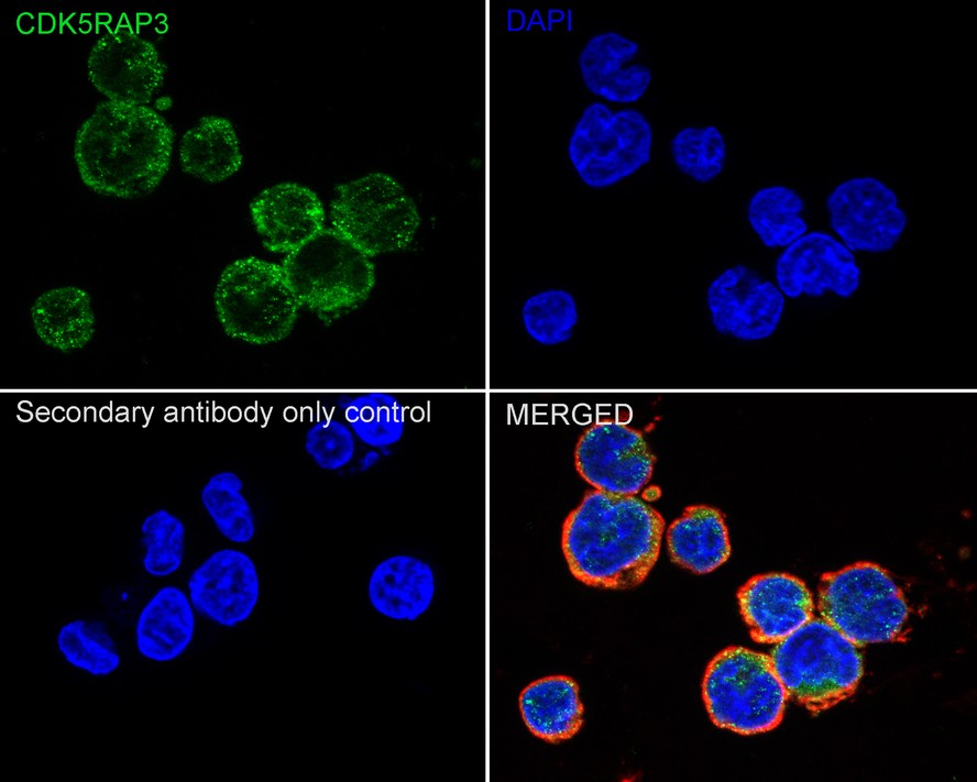
Fig3:
Immunocytochemistry analysis of PC-12 cells labeling CDK5RAP3 with Rabbit anti-CDK5RAP3 antibody (ER1902-72) at 1/100 dilution. Cells were fixed in 4% paraformaldehyde for 15 minutes at room temperature, permeabilized with 0.1% Triton X-100 in PBS for 15 minutes at room temperature, then blocked with 1% BSA in 10% negative goat serum for 1 hour at room temperature. Cells were then incubated with Rabbit anti-CDK5RAP3 antibody (ER1902-72) at 1/100 dilution in 1% BSA in PBST overnight at 4 ℃. Goat Anti-Rabbit IgG H&L (iFluor™ 488, HA1121) was used as the secondary antibody at 1/1,000 dilution. PBS instead of the primary antibody was used as the secondary antibody only control. Nuclear DNA was labelled in blue with DAPI. Beta tubulin (HA601187, red) was stained at 1/100 dilution overnight at +4℃. Goat Anti-Mouse IgG H&L (iFluor™ 594, HA1126) was used as the secondary antibody at 1/1,000 dilution. |
|
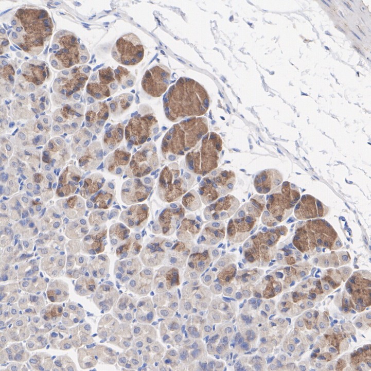
Fig4:
Immunohistochemical analysis of paraffin-embedded mouse stomach tissue with Rabbit anti-CDK5RAP3 antibody (ER1902-72) at 1/200 dilution. The section was pre-treated using heat mediated antigen retrieval with Tris-EDTA buffer (pH 9.0) for 20 minutes. The tissues were blocked in 1% BSA for 20 minutes at room temperature, washed with ddH2O and PBS, and then probed with the primary antibody (ER1902-72) at 1/200 dilution for 1 hour at room temperature. The detection was performed using an HRP conjugated compact polymer system. DAB was used as the chromogen. Tissues were counterstained with hematoxylin and mounted with DPX. |
|
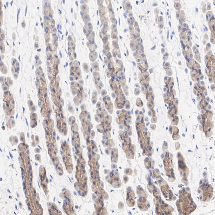
Fig5:
Immunohistochemical analysis of paraffin-embedded rat stomach tissue with Rabbit anti-CDK5RAP3 antibody (ER1902-72) at 1/200 dilution. The section was pre-treated using heat mediated antigen retrieval with Tris-EDTA buffer (pH 9.0) for 20 minutes. The tissues were blocked in 1% BSA for 20 minutes at room temperature, washed with ddH2O and PBS, and then probed with the primary antibody (ER1902-72) at 1/200 dilution for 1 hour at room temperature. The detection was performed using an HRP conjugated compact polymer system. DAB was used as the chromogen. Tissues were counterstained with hematoxylin and mounted with DPX. |
Note: All products are “FOR RESEARCH USE ONLY AND ARE NOT INTENDED FOR DIAGNOSTIC OR THERAPEUTIC USE”.