CD68 Rabbit Polyclonal Antibody

cat.: ER1902-74
| Product Type: | Rabbit polyclonal IgG, primary antibodies |
|---|---|
| Species reactivity: | Human, Mouse |
| Applications: | WB, FC |
| Clonality: | Polyclonal |
| Form: | Liquid |
| Storage condition: | Store at +4℃ after thawing. Aliquot store at -20℃. Avoid repeated freeze / thaw cycles. |
| Storage buffer: | 1*PBS (pH7.4), 0.2% BSA, 50% Glycerol. Preservative: 0.05% Sodium Azide. |
| Concentration: | 1ug/ul |
| Purification: | Immunogen affinity purified. |
| Molecular weight: | Predicted band size 37 kDa. |
| Isotype: | IgG |
| Immunogen: | Synthetic peptide within human CD68 aa 50-120. |
| Positive control: | MCF-7 cell lysate, human lung tissue lysate, THP-1, mouse skin tissue lysate, mouse lung tissue lysate, mouse liver tissue lysate, mouse brain tissue lysate, RAW264.7 cell lysate, NIH/3T3 cell lysate. |
| Subcellular location: | Cell membrane, Endosome, Lysosome, Membrane |
| Recommended Dilutions:
WB FC |
1:1,000 1:50-1:100 |
| Uniprot #: | SwissProt: P34810 Human | P31996 Mouse |
| Alternative names: | CD 68 CD68 CD68 antigen CD68 molecule CD68_HUMAN DKFZp686M18236 gp11 Gp110 LAMP4 Macrophage antigen CD68 (microsialin) MACROPHAGE ANTIGEN CD68 Macrosialin SCARD1 Scavenger receptor class D member 1 |
Images
|
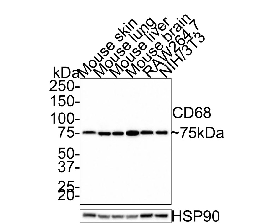
Fig1:
Western blot analysis of CD68 on different lysates with Rabbit anti-CD68 antibody (ER1902-74) at 1/1,000 dilution. Lane 1: Mouse skin tissue lysate (30 µg/Lane) Lane 2: Mouse lung tissue lysate (30 µg/Lane) Lane 3: Mouse liver tissue lysate (30 µg/Lane) Lane 4: Mouse brain tissue lysate (30 µg/Lane) Lane 5: RAW264.7 cell lysate (15 µg/Lane) Lane 6: NIH/3T3 cell lysate (15 µg/Lane) Predicted band size: 37 kDa Observed band size: 75 kDa Exposure time: 2 minutes; 4-20% SDS-PAGE gel. Proteins were transferred to a PVDF membrane and blocked with 5% NFDM/TBST for 1 hour at room temperature. The primary antibody (ER1902-74) at 1/1,000 dilution was used in 5% NFDM/TBST at room temperature for 2 hours. Goat Anti-Rabbit IgG - HRP Secondary Antibody (HA1001) at 1:200,000 dilution was used for 1 hour at room temperature. |
|
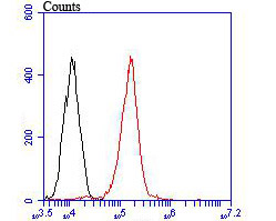
Fig2: Flow cytometric analysis of CD68 was done on THP-1 cells. The cells were fixed, permeabilized and stained with the primary antibody (ER1902-74, 1/50) (red). After incubation of the primary antibody at room temperature for an hour, the cells were stained with a Alexa Fluor®488 conjugate-Goat anti-Rabbit IgG Secondary antibody at 1/1000 dilution for 30 minutes.Unlabelled sample was used as a control (cells without incubation with primary antibody; black). |
Note: All products are “FOR RESEARCH USE ONLY AND ARE NOT INTENDED FOR DIAGNOSTIC OR THERAPEUTIC USE”.