Thymidine Phosphorylase Rabbit Polyclonal Antibody

cat.: ER1902-79
| Product Type: | Rabbit polyclonal IgG, primary antibodies |
|---|---|
| Species reactivity: | Human |
| Applications: | WB, IHC-P, FC |
| Clonality: | Polyclonal |
| Form: | Liquid |
| Storage condition: | Shipped at 4℃. Store at +4℃ short term (1-2 weeks). It is recommended to aliquot into single-use upon delivery. Store at -20℃ long term. |
| Storage buffer: | 1*PBS (pH7.4), 0.2% BSA, 50% Glycerol. Preservative: 0.05% Sodium Azide. |
| Concentration: | 1ug/ul |
| Purification: | Immunogen affinity purified. |
| Molecular weight: | Predicted band size 50 kDa. |
| Isotype: | IgG |
| Immunogen: | Recombinant protein within Human Thymidine Phosphorylase aa 19-239 / 482. |
| Positive control: | SKBR-3 cell lysates, human appendix tissue, human tonsil tissue, human liver cancer tissue, Siha. |
| Subcellular location: | Cytosol. |
| Recommended Dilutions:
WB IHC-P FC |
1:500-1:2,000 1:50-1:400 1:100-1:200 |
| Uniprot #: | SwissProt: P19971 Human |
| Alternative names: | ECGF 1 ECGF ECGF1 Endothelial cell growth factor 1 Endothelial cell growth factor 1 platelet derived Endothelial cell growth factor, platelet-derived Gliostatin hPD ECGF MEDPS1 MNGIE MTDPS1 PD ECGF PD-ECGF PDECGF PDEGF Platelet derived endothelial cell growth factor Platelet derived endothelial growth factor Platelet-derived endothelial cell growth factor TdRPase Thymidine phosphorylase TP Tymp TYPH_HUMAN |
Images
|
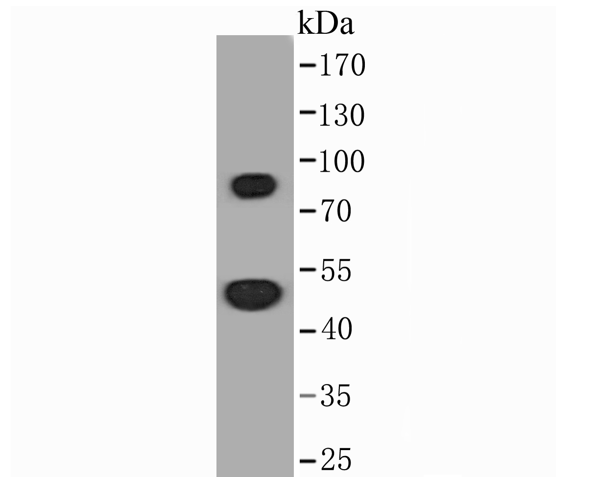
Fig1: Western blot analysis of Thymidine Phosphorylase on SKBR-3 cell lysate. Proteins were transferred to a PVDF membrane and blocked with 5% BSA in PBS for 1 hour at room temperature. The primary antibody (ER1902-79, 1/500) was used in 5% BSA at room temperature for 2 hours. Goat Anti-Rabbit IgG - HRP Secondary Antibody (HA1001) at 1:5,000 dilution was used for 1 hour at room temperature. |
|
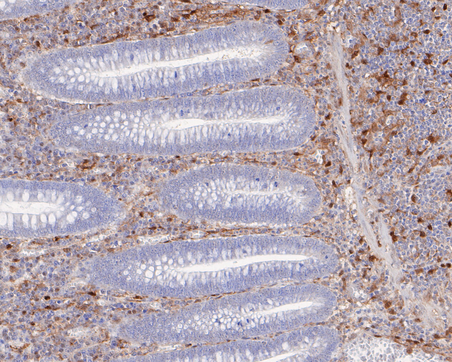
Fig2: Immunohistochemical analysis of paraffin-embedded human appendix tissue using anti-Thymidine Phosphorylase antibody. The section was pre-treated using heat mediated antigen retrieval with Tris-EDTA buffer (pH 8.0-8.4) for 20 minutes.The tissues were blocked in 5% BSA for 30 minutes at room temperature, washed with ddH2O and PBS, and then probed with the primary antibody (ER1902-79, 1/400) for 30 minutes at room temperature. The detection was performed using an HRP conjugated compact polymer system. DAB was used as the chromogen. Tissues were counterstained with hematoxylin and mounted with DPX. |
|
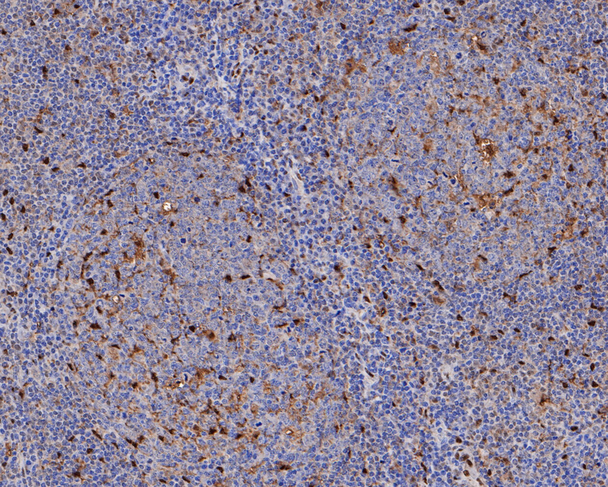
Fig3: Immunohistochemical analysis of paraffin-embedded human tonsil tissue using anti-Thymidine Phosphorylase antibody. The section was pre-treated using heat mediated antigen retrieval with Tris-EDTA buffer (pH 8.0-8.4) for 20 minutes.The tissues were blocked in 5% BSA for 30 minutes at room temperature, washed with ddH2O and PBS, and then probed with the primary antibody (ER1902-79, 1/400) for 30 minutes at room temperature. The detection was performed using an HRP conjugated compact polymer system. DAB was used as the chromogen. Tissues were counterstained with hematoxylin and mounted with DPX. |
|
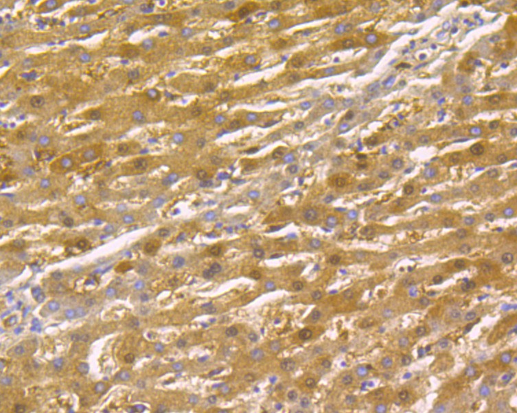
Fig4: Immunohistochemical analysis of paraffin-embedded human liver cancer tissue using anti-Thymidine Phosphorylase antibody. The section was pre-treated using heat mediated antigen retrieval with Tris-EDTA buffer (pH 8.0-8.4) for 20 minutes.The tissues were blocked in 5% BSA for 30 minutes at room temperature, washed with ddH2O and PBS, and then probed with the primary antibody (ER1902-79, 1/200) for 30 minutes at room temperature. The detection was performed using an HRP conjugated compact polymer system. DAB was used as the chromogen. Tissues were counterstained with hematoxylin and mounted with DPX. |
|
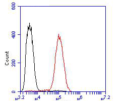
Fig5: Flow cytometric analysis of Thymidine Phosphorylase was done on Siha cells. The cells were fixed, permeabilized and stained with the primary antibody (ER1902-79, 1/100) (red). After incubation of the primary antibody at room temperature for an hour, the cells were stained with a Alexa Fluor 488-conjugated goat anti-rabbit IgG Secondary antibody at 1/500 dilution for 30 minutes.Unlabelled sample was used as a control (cells without incubation with primary antibody; black). |
Note: All products are “FOR RESEARCH USE ONLY AND ARE NOT INTENDED FOR DIAGNOSTIC OR THERAPEUTIC USE”.