TRPM4 Rabbit Polyclonal Antibody

cat.: ER1902-81
| Product Type: | Rabbit polyclonal IgG, primary antibodies |
|---|---|
| Species reactivity: | Human, Mouse, Rat |
| Applications: | WB, FC |
| Clonality: | Polyclonal |
| Form: | Liquid |
| Storage condition: | Shipped at 4℃. Store at +4℃ short term (1-2 weeks). It is recommended to aliquot into single-use upon delivery. Store at -20℃ long term. |
| Storage buffer: | 1*PBS (pH7.4), 0.2% BSA, 50% Glycerol. Preservative: 0.05% Sodium Azide. |
| Concentration: | 1ug/ul |
| Purification: | Immunogen affinity purified. |
| Molecular weight: | Predicted band size 134 kDa. |
| Isotype: | IgG |
| Immunogen: | Synthetic peptide within Human TRPM4 aa 1-50 / 1,214. |
| Positive control: | Mouse colon tissue lysates, MCF-7. |
| Subcellular location: | Cell membrane, endoplasmic reticulum, golgi apparatus. |
| Recommended Dilutions:
WB FC |
1:1,000 1:50-1:100 |
| Uniprot #: | SwissProt: Q8TD43 Human | Q7TN37 Mouse | Q9ESQ5 Rat |
| Alternative names: | 1110030C19Rik AW047689 Calcium-activated non-selective cation channel 1 FLJ20041 hTRPM4 Long transient receptor potential channel 4 LTrpC-4 LTrpC4 Melastatin 4 Melastatin like 2 protein Melastatin-4 Melastatin-like 2 Mls2s PFHB1B Transient receptor potential cation channel subfamily M member 4 Transient receptor potential cation channel, subfamily M, member 4 Trpm4 TRPM4_HUMAN TRPM4B |
Images
|
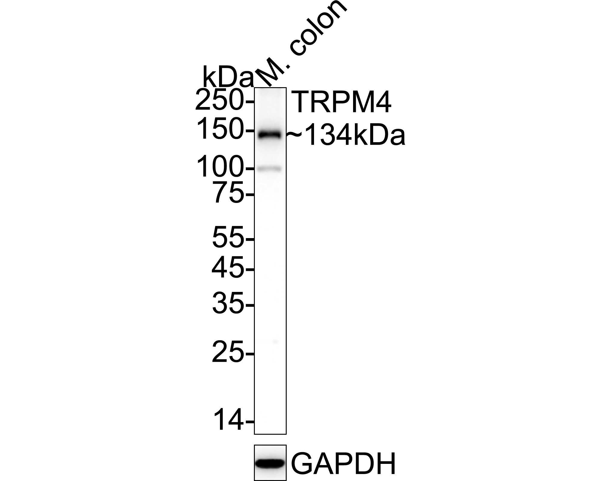
Fig1:
Western blot analysis of TRPM4 on mouse colon tissue lysates with Rabbit anti-TRPM4 antibody (ER1902-81) at 1/1,000 dilution. Lysates/proteins at 40 µg/Lane. Predicted band size: 134 kDa Observed band size: 134 kDa Exposure time: 1 minute 7 seconds; ECL: K1801; 4-20% SDS-PAGE gel. Proteins were transferred to a PVDF membrane and blocked with 5% NFDM/TBST for 1 hour at room temperature. The primary antibody (ER1902-81) at 1/1,000 dilution was used in 5% NFDM/TBST at 4℃ overnight. Goat Anti-Rabbit IgG - HRP Secondary Antibody (HA1001) at 1/50,000 dilution was used for 1 hour at room temperature. |
|
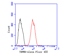
Fig2: Flow cytometric analysis of TRPM4 was done on MCF-7 cells. The cells were fixed, permeabilized and stained with the primary antibody (ER1902-81, 1/50) (red). After incubation of the primary antibody at room temperature for an hour, the cells were stained with a Alexa Fluor 488-conjugated Goat anti-Rabbit IgG Secondary antibody at 1/1000 dilution for 30 minutes.Unlabelled sample was used as a control (cells without incubation with primary antibody; black). |
Note: All products are “FOR RESEARCH USE ONLY AND ARE NOT INTENDED FOR DIAGNOSTIC OR THERAPEUTIC USE”.