P2X3 Rabbit Polyclonal Antibody

cat.: ER1902-88
| Product Type: | Rabbit polyclonal IgG, primary antibodies |
|---|---|
| Species reactivity: | Human, Mouse, Rat |
| Applications: | WB, IHC-P |
| Clonality: | Polyclonal |
| Form: | Liquid |
| Storage condition: | Shipped at 4℃. Store at +4℃ short term (1-2 weeks). It is recommended to aliquot into single-use upon delivery. Store at -20℃ long term. |
| Storage buffer: | 1*PBS (pH7.4), 0.2% BSA, 50% Glycerol. Preservative: 0.05% Sodium Azide. |
| Concentration: | 1ug/ul |
| Purification: | Immunogen affinity purified. |
| Molecular weight: | Predicted band size: 44 kDa |
| Isotype: | IgG |
| Immunogen: | Synthetic peptide within mouse P2X3 aa 348-397 / 397. |
| Positive control: | Mouse testis tissue lysates, rat spinal cord tissue, mouse brain tissue. |
| Subcellular location: | Cell membrane. |
| Recommended Dilutions:
WB IHC-P |
1:500-1:1,000 1:50-1:200 |
| Uniprot #: | SwissProt: P56373 Human | Q3UR32 Mouse | P49654 Rat |
| Alternative names: | ATP receptor MGC129956 P2rx3 P2RX3_HUMAN P2X purinoceptor 3 P2X receptor subunit 3 P2X3 Purinergic receptor Purinergic receptor P2X ligand gated ion channel 3 Purinergic receptor P2X3 Purinoceptor P2X3 |
Images
|
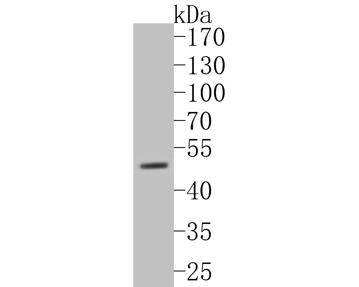
Fig1: Western blot analysis of P2X3 on mouse testis tissue lysates. Proteins were transferred to a PVDF membrane and blocked with 5% BSA in PBS for 1 hour at room temperature. The primary antibody (ER1902-88, 1/500) was used in 5% BSA at room temperature for 2 hours. Goat Anti-Rabbit IgG - HRP Secondary Antibody (HA1001) at 1:5,000 dilution was used for 1 hour at room temperature. |
|
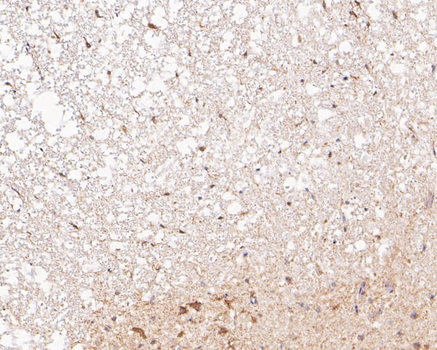
Fig2: Immunohistochemical analysis of paraffin-embedded rat spinal cord tissue using anti-P2X3 antibody. The section was pre-treated using heat mediated antigen retrieval with Tris-EDTA buffer (pH 8.0-8.4) for 20 minutes.The tissues were blocked in 5% BSA for 30 minutes at room temperature, washed with ddH2O and PBS, and then probed with the primary antibody (ER1902-88, 1/200) for 30 minutes at room temperature. The detection was performed using an HRP conjugated compact polymer system. DAB was used as the chromogen. Tissues were counterstained with hematoxylin and mounted with DPX. |
|
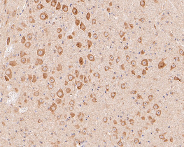
Fig3: Immunohistochemical analysis of paraffin-embedded mouse brain tissue using anti-P2X3 antibody. The section was pre-treated using heat mediated antigen retrieval with Tris-EDTA buffer (pH 8.0-8.4) for 20 minutes.The tissues were blocked in 5% BSA for 30 minutes at room temperature, washed with ddH2O and PBS, and then probed with the primary antibody (ER1902-88, 1/200) for 30 minutes at room temperature. The detection was performed using an HRP conjugated compact polymer system. DAB was used as the chromogen. Tissues were counterstained with hematoxylin and mounted with DPX. |
Note: All products are “FOR RESEARCH USE ONLY AND ARE NOT INTENDED FOR DIAGNOSTIC OR THERAPEUTIC USE”.