FYCO1 Rabbit Polyclonal Antibody
cat.: ER2001-04
Product Type: Rabbit polyclonal IgG, primary antibodies
Species reactivity: Human, Mouse, Rat
Applications: WB, IHC-P, FC
Clonality: Polyclonal
Form: Liquid
Storage condition: Store at +4℃ after thawing. Aliquot store at -20℃. Avoid repeated freeze / thaw cycles.
Storage buffer: 1*TBS (pH7.4), 0.2% BSA, 50% Glycerol. Preservative: 0.05% Sodium Azide.
Concentration: 1ug/ul
Purification: Immunogen affinity purified.
Molecular weight: 167 kDa
Isotype: IgG
Immunogen: Recombinant protein within human FYCO1 aa 1-200.
Positive control: SK-Br-3 cell lysates, rat testis tissue, human liver tissue, human thyroid tissue, human colon tissue, mouse colon tissue, 293.
Subcellular location: Lysosome, Endosome, autophagosome.
Recommended Dilutions:
  WB
  IHC-P
  FC

1:500
1:200-1:1,000
1:50-1:100
Uniprot #: SwissProt: Q9BQS8 Human | Q8VDC1 Mouse
Alternative names: CATC2 CTRCT18 DKFZp779K1152 FLJ13335 FYCO1 FYCO1_HUMAN FYVE and coiled coil domain containing 1 FYVE and coiled coil domain containing protein 1 FYVE and coiled-coil domain-containing protein 1 MGC126517 MGC126519 RUFY3 RUN and FYVE domain containing 3 ZFYVE7 Zinc finger FYVE domain containing protein 7 Zinc finger FYVE domain-containing protein 7
Images
ER2001-04_1.jpg 
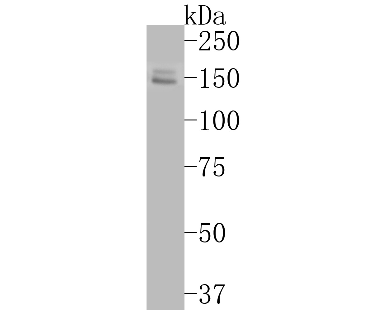
Fig1: Western blot analysis of FYCO1 on SK-Br-3 cell lysates. Proteins were transferred to a PVDF membrane and blocked with 5% BSA in PBS for 1 hour at room temperature. The primary antibody (ER2001-04, 1/500) was used in 5% BSA at room temperature for 2 hours. Goat Anti-Rabbit IgG - HRP Secondary Antibody (HA1001) at 1:5,000 dilution was used for 1 hour at room temperature.
ER2001-04_2.jpg Fig2: Immunohistochemical analysis of paraffin-embedded rat testis tissue using anti-FYCO1 antibody. The section was pre-treated using heat mediated antigen retrieval with Tris-EDTA buffer (pH 8.0-8.4) for 20 minutes.The tissues were blocked in 5% BSA for 30 minutes at room temperature, washed with ddH2O and PBS, and then probed with the primary antibody (ER2001-04, 1/500) for 30 minutes at room temperature. The detection was performed using an HRP conjugated compact polymer system. DAB was used as the chromogen. Tissues were counterstained with hematoxylin and mounted with DPX.
ER2001-04_3.jpg Fig3: Immunohistochemical analysis of paraffin-embedded human liver tissue using anti-FYCO1 antibody. The section was pre-treated using heat mediated antigen retrieval with Tris-EDTA buffer (pH 8.0-8.4) for 20 minutes.The tissues were blocked in 5% BSA for 30 minutes at room temperature, washed with ddH2O and PBS, and then probed with the primary antibody (ER2001-04, 1/500) for 30 minutes at room temperature. The detection was performed using an HRP conjugated compact polymer system. DAB was used as the chromogen. Tissues were counterstained with hematoxylin and mounted with DPX.
ER2001-04_4.jpg Fig4: Immunohistochemical analysis of paraffin-embedded human thyroid tissue using anti-FYCO1 antibody. The section was pre-treated using heat mediated antigen retrieval with Tris-EDTA buffer (pH 8.0-8.4) for 20 minutes.The tissues were blocked in 5% BSA for 30 minutes at room temperature, washed with ddH2O and PBS, and then probed with the primary antibody (ER2001-04, 1/500) for 30 minutes at room temperature. The detection was performed using an HRP conjugated compact polymer system. DAB was used as the chromogen. Tissues were counterstained with hematoxylin and mounted with DPX.
ER2001-04_5.jpg Fig5: Immunohistochemical analysis of paraffin-embedded human colon tissue using anti-FYCO1 antibody. The section was pre-treated using heat mediated antigen retrieval with Tris-EDTA buffer (pH 8.0-8.4) for 20 minutes.The tissues were blocked in 5% BSA for 30 minutes at room temperature, washed with ddH2O and PBS, and then probed with the primary antibody (ER2001-04, 1/500) for 30 minutes at room temperature. The detection was performed using an HRP conjugated compact polymer system. DAB was used as the chromogen. Tissues were counterstained with hematoxylin and mounted with DPX.
ER2001-04_6.jpg Fig6: Immunohistochemical analysis of paraffin-embedded mouse colon tissue using anti-FYCO1 antibody. The section was pre-treated using heat mediated antigen retrieval with Tris-EDTA buffer (pH 8.0-8.4) for 20 minutes.The tissues were blocked in 5% BSA for 30 minutes at room temperature, washed with ddH2O and PBS, and then probed with the primary antibody (ER2001-04, 1/500) for 30 minutes at room temperature. The detection was performed using an HRP conjugated compact polymer system. DAB was used as the chromogen. Tissues were counterstained with hematoxylin and mounted with DPX.
ER2001-04_7.jpg Fig7: Flow cytometric analysis of FYCO1 was done on 293 cells. The cells were fixed, permeabilized and stained with the primary antibody (ER2001-04, 1/50) (red). After incubation of the primary antibody at room temperature for an hour, the cells were stained with a Alexa Fluor 488-conjugated Goat anti-Rabbit IgG Secondary antibody at 1/1000 dilution for 30 minutes.Unlabelled sample was used as a control (cells without incubation with primary antibody; black).
Note: All products are “FOR RESEARCH USE ONLY AND ARE NOT INTENDED FOR DIAGNOSTIC OR THERAPEUTIC USE”.