Androgen Receptor Rabbit Polyclonal Antibody

cat.: ER2001-34
| Product Type: | Rabbit polyclonal IgG, primary antibodies |
|---|---|
| Species reactivity: | Human, Rat |
| Applications: | WB, IF-Cell, FC |
| Clonality: | Polyclonal |
| Form: | Liquid |
| Storage condition: | Store at +4℃ after thawing. Aliquot store at -20℃. Avoid repeated freeze / thaw cycles. |
| Storage buffer: | 1*TBS (pH7.4), 0.2% BSA, 50% Glycerol. Preservative: 0.05% Sodium Azide. |
| Concentration: | 1ug/ul |
| Purification: | Protein A affinity purified. |
| Molecular weight: | 99 kDa |
| Isotype: | IgG |
| Immunogen: | Recombinant protein within human Androgen Receptor aa 200-500. |
| Positive control: | Human skeletal muscle tissue lysate, rat heart tissue lysate, A549, MCF-7. |
| Subcellular location: | Cytoplasm, Nucleus |
| Recommended Dilutions:
WB IF-Cell FC |
1:500-1:1,000 1:50-1:200 1:50-1:100 |
| Uniprot #: | SwissProt: P10275 Human | P15207 Rat |
| Alternative names: | AIS ANDR_HUMAN Androgen nuclear receptor variant 2 Androgen receptor (dihydrotestosterone receptor; testicular feminization; spinal and bulbar muscular atrophy; Kennedy disease) Androgen receptor androgen receptor splice variant 4b AR AR8 DHTR Dihydro testosterone receptor Dihydrotestosterone receptor (DHTR) Dihydrotestosterone receptor HUMARA HYSP1 KD Kennedy disease (KD) NR3C4 Nuclear receptor subfamily 3 group C member 4 (NR3C4) Nuclear receptor subfamily 3 group C member 4 SBMA SMAX1 Spinal and bulbar muscular atrophy (SBMA) Spinal and bulbar muscular atrophy Testicular Feminization (TFM) TFM |
Images
|
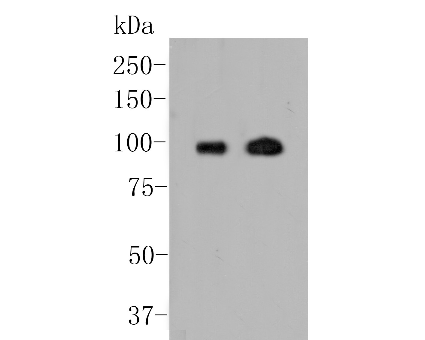
Fig1:
Western blot analysis of Androgen Receptor on different lysates. Proteins were transferred to a PVDF membrane and blocked with 5% BSA in PBS for 1 hour at room temperature. The primary antibody (ER2001-34, 1/500) was used in 5% BSA at room temperature for 2 hours. Goat Anti-Rabbit IgG - HRP Secondary Antibody (HA1001) at 1:5,000 dilution was used for 1 hour at room temperature. Positive control: Lane 1: Human skeletal muscle tissue lysate Lane 2: Rat heart tissue lysate |
|
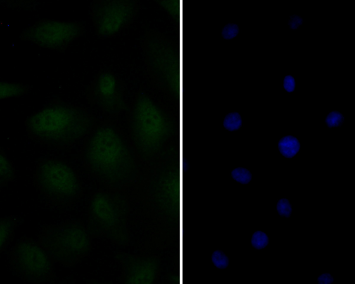
Fig2: ICC staining of Androgen Receptor in A549 cells (green). Formalin fixed cells were permeabilized with 0.1% Triton X-100 in TBS for 10 minutes at room temperature and blocked with 1% Blocker BSA for 15 minutes at room temperature. Cells were probed with the primary antibody (ER2001-34, 1/50) for 1 hour at room temperature, washed with PBS. Alexa Fluor®488 Goat anti-Rabbit IgG was used as the secondary antibody at 1/100 dilution. The nuclear counter stain is DAPI (blue). |
|
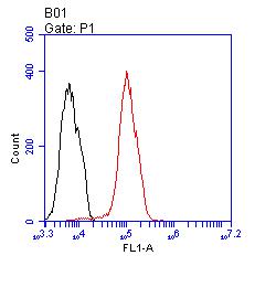
Fig3: Flow cytometric analysis of Androgen Receptor was done on MCF-7 cells. The cells were fixed, permeabilized and stained with the primary antibody (ER2001-34, 1/100) (red). After incubation of the primary antibody at room temperature for an hour, the cells were stained with a Alexa Fluor 488-conjugated goat anti-rabbit IgG Secondary antibody at 1/500 dilution for 30 minutes.Unlabelled sample was used as a control (cells without incubation with primary antibody; black). |
Note: All products are “FOR RESEARCH USE ONLY AND ARE NOT INTENDED FOR DIAGNOSTIC OR THERAPEUTIC USE”.