Nucleoporin p62 Rabbit Polyclonal Antibody

cat.: ER2001-57
| Product Type: | Rabbit polyclonal IgG, primary antibodies |
|---|---|
| Species reactivity: | Human |
| Applications: | WB, IHC-P |
| Clonality: | Polyclonal |
| Form: | Liquid |
| Storage condition: | Shipped at 4℃. Store at +4℃ short term (1-2 weeks). It is recommended to aliquot into single-use upon delivery. Store at -20℃ long term. |
| Storage buffer: | 1*TBS (pH7.4), 0.2% BSA, 50% Glycerol. Preservative: 0.05% Sodium Azide. |
| Concentration: | 1ug/ul |
| Purification: | Immunogen affinity purified. |
| Molecular weight: | Predicted band size: 53 kDa. |
| Isotype: | IgG |
| Immunogen: | Synthetic peptide within human Nucleoporin p62 aa 470-522. |
| Positive control: | K562 cell lysate, A431 cell lysate, human breast carcinoma tissue, human rectum tissue. |
| Subcellular location: | Cytoplasm, Cytoskeleton, Nuclear pore complex, Nucleus. |
| Recommended Dilutions:
WB IHC-P |
1:500-1:1,000 1:100-1:400 |
| Uniprot #: | SwissProt: P37198 Human |
| Alternative names: | 62 kDa nucleoporin DKFZp547L134 FLJ20822 FLJ43869 IBSN MGC841 Nuclear pore glycoprotein p62 nucleoporin 62kDa Nucleoporin Nup62 nucleoporin p62 nucleoporin p62KD NUP62 NUP62 protein p62 SNDI |
Images
|
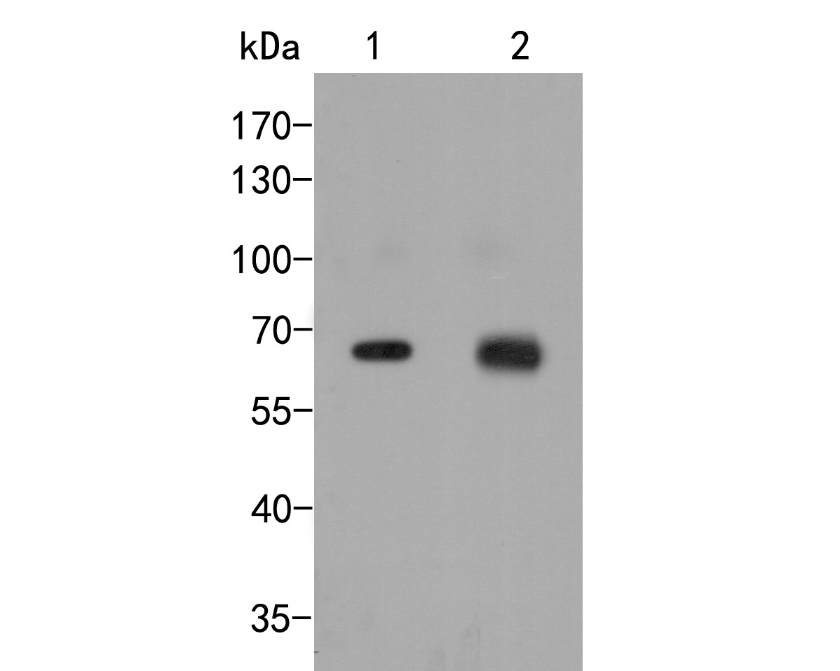
Fig1:
Western blot analysis of Nucleoporin p62 on different lysates. Proteins were transferred to a PVDF membrane and blocked with 5% BSA in PBS for 1 hour at room temperature. The primary antibody (ER2001-57, 1/500) was used in 5% BSA at room temperature for 2 hours. Goat Anti-Rabbit IgG - HRP Secondary Antibody (HA1001) at 1:5,000 dilution was used for 1 hour at room temperature. Positive control: Lane 1: K562 cell lysate Lane 2: A431 cell lysate |
|
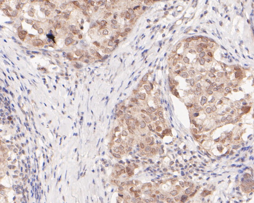
Fig2: Immunohistochemical analysis of paraffin-embedded human breast carcinoma tissue using anti-Nucleoporin p62 antibody. The section was pre-treated using heat mediated antigen retrieval with sodium citrate buffer (pH 6.0) for 20 minutes. The tissues were blocked in 5% BSA for 30 minutes at room temperature, washed with ddH2O and PBS, and then probed with the primary antibody (ER2001-57, 1/400) for 30 minutes at room temperature. The detection was performed using an HRP conjugated compact polymer system. DAB was used as the chromogen. Tissues were counterstained with hematoxylin and mounted with DPX. |
|
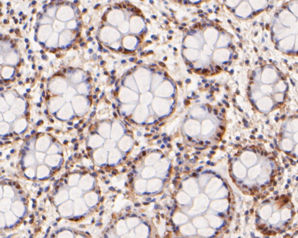
Fig3: Immunohistochemical analysis of paraffin-embedded human rectum tissue using anti-Nucleoporin p62 antibody. The section was pre-treated using heat mediated antigen retrieval with sodium citrate buffer (pH 6.0) for 20 minutes. The tissues were blocked in 5% BSA for 30 minutes at room temperature, washed with ddH2O and PBS, and then probed with the primary antibody (ER2001-57, 1/100) for 30 minutes at room temperature. The detection was performed using an HRP conjugated compact polymer system. DAB was used as the chromogen. Tissues were counterstained with hematoxylin and mounted with DPX. |
Note: All products are “FOR RESEARCH USE ONLY AND ARE NOT INTENDED FOR DIAGNOSTIC OR THERAPEUTIC USE”.