GRP78 / BIP Rabbit Polyclonal Antibody

cat.: ER40402
| Product Type: | Rabbit polyclonal IgG, primary antibodies |
|---|---|
| Species reactivity: | Human, Mouse, Rat |
| Applications: | WB, IF-Cell, IHC-P, FC |
| Clonality: | Polyclonal |
| Form: | Liquid |
| Storage condition: | Store at +4℃ after thawing. Aliquot store at -20℃ or -80℃. Avoid repeated freeze / thaw cycles. |
| Storage buffer: | 1*PBS (pH7.4), 0.2% BSA, 40% Glycerol. Preservative: 0.05% Sodium Azide. |
| Concentration: | 1ug/ul |
| Purification: | Immunogen affinity purified. |
| Molecular weight: | Predicted band size: 72 kDa |
| Isotype: | IgG |
| Immunogen: | Synthetic peptide within C-terminal human GRP78. |
| Positive control: | Hela cell lysate, MCF-7 cell lysate, A549 cell lysate, mouse heart tissue lysate, rat brain tissue lysate, SK-Br-3, HepG2, Hela, rat small intestine tissue, human breast carcinoma tissue. |
| Subcellular location: | Cytoplasm, Endoplasmic reticulum. |
| Recommended Dilutions:
WB IF-Cell IHC-P FC |
1:500-1:2,000 1:50-1:200 1:50-1:200 1:100-1:200 |
| Uniprot #: | SwissProt: P11021 Human | P20029 Mouse | P06761 Rat |
| Alternative names: | 78 kDa glucose regulated protein 78 kDa glucose-regulated protein AL022860 AU019543 BIP D2Wsu141e D2Wsu17e Endoplasmic reticulum lumenal Ca(2+)-binding protein grp78 Endoplasmic reticulum lumenal Ca2+ binding protein grp78 Epididymis secretory sperm binding protein Li 89n FLJ26106 Glucose Regulated Protein 78kDa GRP 78 GRP-78 GRP78 GRP78_HUMAN Heat shock 70 kDa protein 5 Heat Shock 70kDa Protein 5 Heat shock protein family A (Hsp70) member 5 HEL S 89n Hsce70 HSPA 5 HSPA5 Immunoglobulin Heavy Chain Binding Protein Immunoglobulin heavy chain-binding protein mBiP MIF2 Sez7 |
Images
|
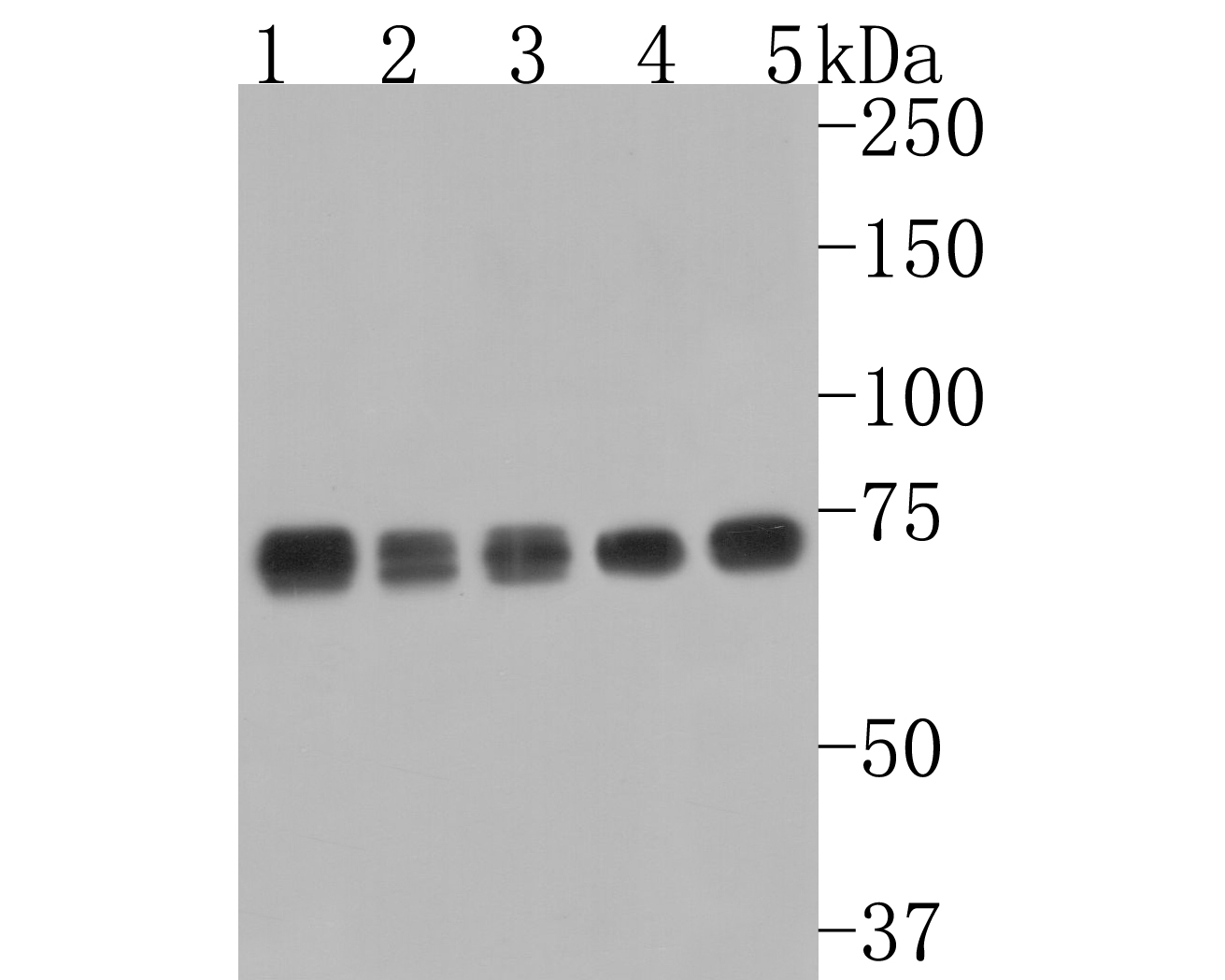
Fig1:
Western blot analysis of GRP78 / BIP on different lysates. Proteins were transferred to a PVDF membrane and blocked with 5% BSA in PBS for 1 hour at room temperature. The primary antibody (ER40402, 1/1,000) was used in 5% BSA at room temperature for 2 hours. Goat Anti-Rabbit IgG - HRP Secondary Antibody (HA1001) at 1:200,000 dilution was used for 1 hour at room temperature. Positive control: Lane 1: Hela cell lysate Lane 2: MCF-7 cell lysate Lane 3: A549 cell lysate Lane 4: Mouse heart tissue lysate Lane 5: Rat brain tissue lysate |
|
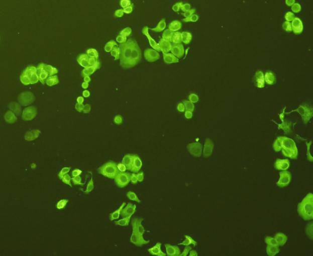
Fig2: ICC staining of GRP78 / BIP in SK-Br-3 cells (green). Formalin fixed cells were permeabilized with 0.1% Triton X-100 in TBS for 10 minutes at room temperature and blocked with 10% negative goat serum for 15 minutes at room temperature. Cells were probed with the primary antibody (ER40402, 1/50) for 1 hour at room temperature, washed with PBS. Alexa Fluor®488 conjugate-Goat anti-Rabbit IgG was used as the secondary antibody at 1/1,000 dilution. The nuclear counter stain is DAPI (blue). |
|
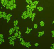
Fig3: ICC staining of GRP78 / BIP in HepG2 cells (green). Formalin fixed cells were permeabilized with 0.1% Triton X-100 in TBS for 10 minutes at room temperature and blocked with 10% negative goat serum for 15 minutes at room temperature. Cells were probed with the primary antibody (ER40402, 1/50) for 1 hour at room temperature, washed with PBS. Alexa Fluor®488 conjugate-Goat anti-Rabbit IgG was used as the secondary antibody at 1/1,000 dilution. The nuclear counter stain is DAPI (blue). |
|
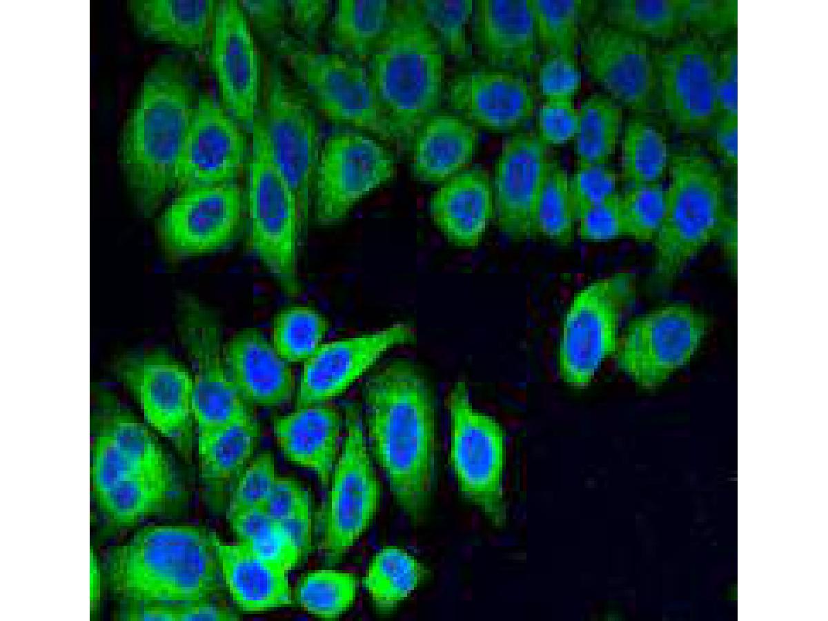
Fig4: ICC staining of GRP78 / BIP in Hela cells (green). Formalin fixed cells were permeabilized with 0.1% Triton X-100 in TBS for 10 minutes at room temperature and blocked with 10% negative goat serum for 15 minutes at room temperature. Cells were probed with the primary antibody (ER40402, 1/50) for 1 hour at room temperature, washed with PBS. Alexa Fluor®488 conjugate-Goat anti-Rabbit IgG was used as the secondary antibody at 1/1,000 dilution. The nuclear counter stain is DAPI (blue). |
|
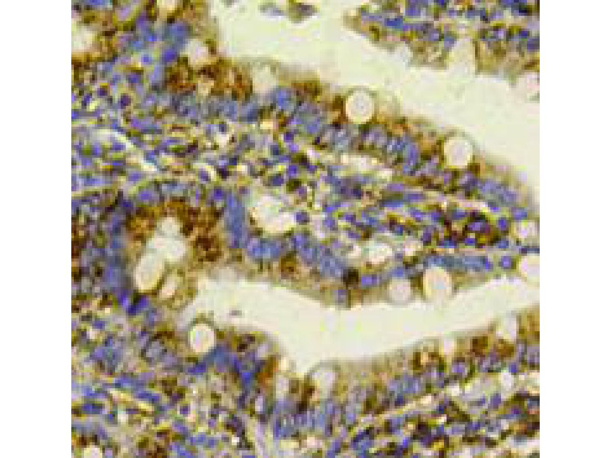
Fig5: Immunohistochemical analysis of paraffin-embedded rat small intestine tissue using anti-GRP78 / BIP antibody. The section was pre-treated using heat mediated antigen retrieval with Tris-EDTA buffer (pH 9.0) for 20 minutes.The tissues were blocked in 1% BSA for 30 minutes at room temperature, washed with ddH2O and PBS, and then probed with the primary antibody (ER40402, 1/50) for 30 minutes at room temperature. The detection was performed using an HRP conjugated compact polymer system. DAB was used as the chromogen. Tissues were counterstained with hematoxylin and mounted with DPX. |
|
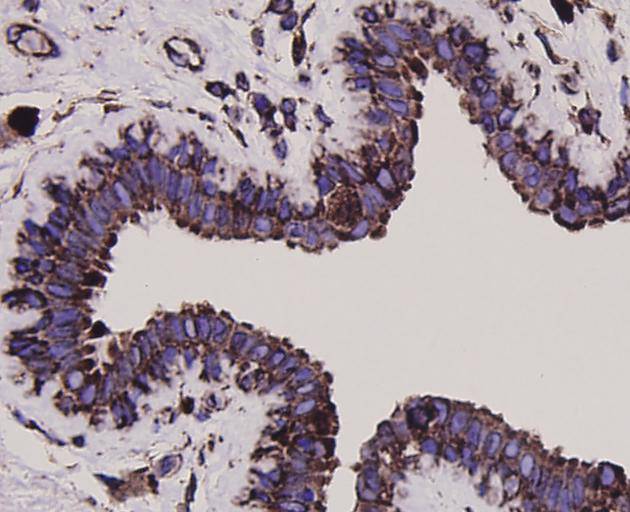
Fig6: Immunohistochemical analysis of paraffin-embedded human breast carcinoma tissue using anti-GRP78 / BIP antibody. The section was pre-treated using heat mediated antigen retrieval with Tris-EDTA buffer (pH 9.0) for 20 minutes.The tissues were blocked in 1% BSA for 30 minutes at room temperature, washed with ddH2O and PBS, and then probed with the primary antibody (ER40402, 1/50) for 30 minutes at room temperature. The detection was performed using an HRP conjugated compact polymer system. DAB was used as the chromogen. Tissues were counterstained with hematoxylin and mounted with DPX. |
Note: All products are “FOR RESEARCH USE ONLY AND ARE NOT INTENDED FOR DIAGNOSTIC OR THERAPEUTIC USE”.