ERG Recombinant Rabbit Monoclonal Antibody [SP06-04]

cat.: ET1604-21
| Product Type: | Recombinant Rabbit monoclonal IgG, primary antibodies |
|---|---|
| Species reactivity: | Human, Mouse, Rat |
| Applications: | WB, IF-Tissue, IHC-P |
| Clonality: | Monoclonal |
| Clone number: | SP06-04 |
| Form: | Liquid |
| Storage condition: | Shipped at 4℃. Store at +4℃ short term (1-2 weeks). Store at -20℃ long term. |
| Storage buffer: | 1*TBS (pH7.4), 0.05% BSA, 40% Glycerol. Preservative: 0.05% Sodium Azide. |
| Concentration: | 1ug/ul |
| Purification: | Protein A affinity purified. |
| Molecular weight: | 54 kDa |
| Isotype: | IgG |
| Immunogen: | Synthetic peptide within Human ERG aa 430-479 / 479. |
| Positive control: | Jurkat cell lysates, CRC, PC-3M, human colon carcinoma tissue, mouse brain tissue, mouse heart tissue, human spleen tissue, human kidney tissue. |
| Subcellular location: | Cytoplasm, Nucleus. |
| Recommended Dilutions:
WB IF-Tissue IHC-P |
1:2,000-1:5,000 1:200-1:500 1:200-1:1,000 |
| Uniprot #: | SwissProt: P11308 Human | P81270 Mouse Unigene: 47987 Rat |
| Alternative names: | Avian erythroblastosis virus E-26 (v-ets) oncogene related D030036I24Rik Erg 3 Erg ERG/EWS fusion gene, included ERG/FUS fusion gene, included ERG/TMPSSR2 fusion gene, included ERG_HUMAN ERG1, included ERG2, included ets related ETS-related gene KCNH2 Oncogene ERG p55 TMPRSS2/ERG fusion transcriptional regulator ERG (transforming protein ERG) Transcriptional regulator ERG Transforming protein ERG v ets avian erythroblastosis virus E26 oncogene v ets avian erythroblastosis virus E26 oncogene related v ets erythroblastosis virus E26 oncogene homolog v ets erythroblastosis virus E26 oncogene like v ets erythroblastosis virus E26 oncogene like isoform 2 v-ets erythroblastosis virus E26 oncogene v-ets erythroblastosis virus E26 oncogene homolog (avian) V-ets erythroblastosis virus E26 oncogene like (Avian), isoform CRA_e |
Images

|
Fig1:
Western blot analysis of ERG on Jurkat cell lysates with Rabbit anti-ERG antibody (ET1604-21) at 1/2,000 dilution. Lysates/proteins at 20 µg/Lane. Predicted band size: 54 kDa Observed band size: 54 kDa Exposure time: 20 seconds; 4-20% SDS-PAGE gel. Proteins were transferred to a PVDF membrane and blocked with 5% NFDM/TBST for 1 hour at room temperature. The primary antibody (ET1604-21) at 1/2,000 dilution was used in 5% NFDM/TBST at 4℃ overnight. Goat Anti-Rabbit IgG - HRP Secondary Antibody (HA1001) at 1/50,000 dilution was used for 1 hour at room temperature. |

|
Fig2:
Western blot analysis of ERG on different lysates with Rabbit anti-ERG antibody (ET1604-21) at 1/2,000 dilution. Lane 1: Mouse lung tissue lysate Lane 2: Mouse spleen tissue lysate Lane 3: Rat brain tissue lysate Lane 4: Rat lung tissue lysate Lysates/proteins at 20 µg/Lane. Predicted band size: 54 kDa Observed band size: 54 kDa Exposure time: 1 minutes 2 seconds; 4-20% SDS-PAGE gel. Proteins were transferred to a PVDF membrane and blocked with 5% NFDM/TBST for 1 hour at room temperature. The primary antibody (ET1604-21) at 1/2,000 dilution was used in 5% NFDM/TBST at 4℃ overnight. Goat Anti-Rabbit IgG - HRP Secondary Antibody (HA1001) at 1/50,000 dilution was used for 1 hour at room temperature. |

|
Fig3:
Immunohistochemical analysis of paraffin-embedded human spleen tissue with Rabbit anti-ERG antibody (ET1604-21) at 1/200 dilution. The section was pre-treated using heat mediated antigen retrieval with sodium citrate buffer (pH 6.0) (high pressure) for 2 minutes. The tissues were blocked in 1% BSA for 20 minutes at room temperature, washed with ddH2O and PBS, and then probed with the primary antibody (ET1604-21) at 1/200 dilution for 1 hour at room temperature. The detection was performed using an HRP conjugated compact polymer system. DAB was used as the chromogen. Tissues were counterstained with hematoxylin and mounted with DPX. |

|
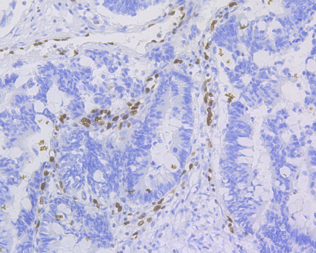
Fig4:
Immunohistochemical analysis of paraffin-embedded human prostate cancer tissue with Rabbit anti-ERG antibody (ET1604-21) at 1/500 dilution. The section was pre-treated using heat mediated antigen retrieval with sodium citrate buffer (pH 6.0) (high pressure) for 2 minutes. The tissues were blocked in 1% BSA for 20 minutes at room temperature, washed with ddH2O and PBS, and then probed with the primary antibody (ET1604-21) at 1/100 dilution for 1 hour at room temperature. The detection was performed using an HRP conjugated compact polymer system. DAB was used as the chromogen. Tissues were counterstained with hematoxylin and mounted with DPX. |
|
Fig5:
Immunohistochemical analysis of paraffin-embedded mouse brain tissue using anti-ERG antibody. The section was pre-treated using heat mediated antigen retrieval with sodium citrate buffer (pH 6.0) for 20 minutes. The tissues were blocked in 5% BSA for 30 minutes at room temperature, washed with ddH2O and PBS, and then probed with the primary antibody (ET1604-21, 1/100) for 30 minutes at room temperature. The detection was performed using an HRP conjugated compact polymer system. DAB was used as the chromogen. Tissues were counterstained with hematoxylin and mounted with DPX. |

|
Fig6:
Immunohistochemical analysis of paraffin-embedded mouse brain tissue with Rabbit anti-ERG antibody (ET1604-21) at 1/1,000 dilution. The section was pre-treated using heat mediated antigen retrieval with sodium citrate buffer (pH 6.0) (high pressure) for 2 minutes. The tissues were blocked in 1% BSA for 20 minutes at room temperature, washed with ddH2O and PBS, and then probed with the primary antibody (ET1604-21) at 1/1,000 dilution for 1 hour at room temperature. The detection was performed using an HRP conjugated compact polymer system. DAB was used as the chromogen. Tissues were counterstained with hematoxylin and mounted with DPX. |

|
Fig7:
Immunohistochemical analysis of paraffin-embedded mouse hippocampus tissue with Rabbit anti-ERG antibody (ET1604-21) at 1/1,000 dilution. The section was pre-treated using heat mediated antigen retrieval with sodium citrate buffer (pH 6.0) (high pressure) for 2 minutes. The tissues were blocked in 1% BSA for 20 minutes at room temperature, washed with ddH2O and PBS, and then probed with the primary antibody (ET1604-21) at 1/1,000 dilution for 1 hour at room temperature. The detection was performed using an HRP conjugated compact polymer system. DAB was used as the chromogen. Tissues were counterstained with hematoxylin and mounted with DPX. |

|
Fig8:
Immunohistochemical analysis of paraffin-embedded rat brain tissue with Rabbit anti-ERG antibody (ET1604-21) at 1/1,000 dilution. The section was pre-treated using heat mediated antigen retrieval with sodium citrate buffer (pH 6.0) (high pressure) for 2 minutes. The tissues were blocked in 1% BSA for 20 minutes at room temperature, washed with ddH2O and PBS, and then probed with the primary antibody (ET1604-21) at 1/1,000 dilution for 1 hour at room temperature. The detection was performed using an HRP conjugated compact polymer system. DAB was used as the chromogen. Tissues were counterstained with hematoxylin and mounted with DPX. |

|
Fig9:
Immunohistochemical analysis of paraffin-embedded human kidney tissue with Rabbit anti-ERG antibody (ET1604-21) at 1/500 dilution. The section was pre-treated using heat mediated antigen retrieval with sodium citrate buffer (pH 6.0) for 2 minutes. The tissues were blocked in 1% BSA for 20 minutes at room temperature, washed with ddH2O and PBS, and then probed with the primary antibody (ET1604-21) at 1/500 dilution for 1 hour at room temperature. The detection was performed using an HRP conjugated compact polymer system. DAB was used as the chromogen. Tissues were counterstained with hematoxylin and mounted with DPX. |

|
Fig10:
Immunohistochemical analysis of paraffin-embedded human kidney tissue with Rabbit anti-ERG antibody (ET1604-21) at 1/200 dilution. The section was incubated with ET1604-21 for 32 mins at room temperature. The immunostaining was performed on a Roche Benchmark XT instrument. DAB was used as the chromogen. Tissues were counterstained with hematoxylin and mounted with DPX. |

|
Fig11:
Immunohistochemical analysis of paraffin-embedded human kidney tissue with Rabbit anti-ERG antibody (ET1604-21) at 1/200 dilution. Heat mediated antigen retrieval with Tris-EDTA buffer (pH 9.0, epitope retrieval solution 2) for 20 mins. The section was incubated with ET1604-21 for 30 mins at room temperature. The immunostaining was performed on a Leica Biosystems BOND® RX instrument. DAB was used as the chromogen. Tissues were counterstained with hematoxylin and mounted with DPX. |
Note: All products are “FOR RESEARCH USE ONLY AND ARE NOT INTENDED FOR DIAGNOSTIC OR THERAPEUTIC USE”.