CD8 alpha Recombinant Rabbit Monoclonal Antibody [SI18-01]

cat.: ET1606-31
| Product Type: | Recombinant Rabbit monoclonal IgG, primary antibodies |
|---|---|
| Species reactivity: | Human |
| Applications: | WB, IF-Cell, IF-Tissue, IHC-P, FC |
| Clonality: | Monoclonal |
| Clone number: | SI18-01 |
| Form: | Liquid |
| Storage condition: | Shipped at 4℃. Store at +4℃ short term (1-2 weeks). Store at -20℃ long term. |
| Storage buffer: | 1*TBS (pH7.4), 0.05% BSA, 40% Glycerol. Preservative: 0.05% Sodium Azide. |
| Concentration: | 1ug/ul |
| Purification: | Protein A affinity purified. |
| Molecular weight: | Predicted band size: 26 kDa |
| Isotype: | IgG |
| Immunogen: | Synthetic peptide within Human CD8 alpha aa 186-235 / 235. |
| Positive control: | THP-1 cell lysate, Jurkat cell lysate, PC-3M cell lysate, Hela, CRC, human spleen tissue, human lymph node tissue, human appendix tissue, Jurkat. |
| Subcellular location: | Cell membrane, Secreted. |
| Recommended Dilutions:
WB IF-Cell IF-Tissue IHC-P FC |
1:2,000-1:5,000 1:100-1:200 1:100-1:200 1:100-1:200 1:500-1:1,000 |
| Uniprot #: | SwissProt: P01732 Human |
| Alternative names: | alpha polypeptide (p32) CD8 CD8 antigen alpha polypeptide CD8 antigen alpha polypeptide (p32) CD8a CD8A antigen CD8A molecule CD8A_HUMAN Leu2 Leu2 T lymphocyte antigen Ly3 LYT3 MAL OKT8 T cell antigen OTTHUMP00000160760 OTTHUMP00000160764 OTTHUMP00000203528 OTTHUMP00000203721 p32 T cell antigen Leu2 T cell co receptor T-cell surface glycoprotein CD8 alpha chain T-lymphocyte differentiation antigen T8/Leu-2 T8 T cell antigen T8/Leu-2 T-lymphocyte differentiation antigen |
Images

|
Fig1:
Western blot analysis of CD8 alpha on different lysates with Rabbit anti-CD8 alpha antibody (ET1606-31) at 1/2,000 dilution. Lane 1: THP-1 cell lysate Lane 2: Jurkat cell lysate Lane 3: PC-3M cell lysate Lysates/proteins at 20 µg/Lane. Predicted band size: 26 kDa Observed band size: 35 kDa Exposure time: 2 minutes; 4-20% SDS-PAGE gel. Proteins were transferred to a PVDF membrane and blocked with 5% NFDM/TBST for 1 hour at room temperature. The primary antibody (ET1606-31) at 1/2,000 dilution was used in 5% NFDM/TBST at room temperature for 2 hours. Goat Anti-Rabbit IgG - HRP Secondary Antibody (HA1001) at 1:100,000 dilution was used for 1 hour at room temperature. |
|
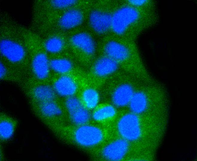
Fig2: ICC staining of CD8 alpha in Hela cells (green). Formalin fixed cells were permeabilized with 0.1% Triton X-100 in TBS for 10 minutes at room temperature and blocked with 1% Blocker BSA for 15 minutes at room temperature. Cells were probed with the primary antibody (ET1606-31, 1/50) for 1 hour at room temperature, washed with PBS. Alexa Fluor®488 Goat anti-Rabbit IgG was used as the secondary antibody at 1/1,000 dilution. The nuclear counter stain is DAPI (blue). |

|
Fig3:
Immunohistochemical analysis of paraffin-embedded human spleen tissue using anti-CD8 alpha antibody. The section was pre-treated using heat mediated antigen retrieval with Tris-EDTA buffer (pH 8.0-8.4) for 20 minutes.The tissues were blocked in 5% BSA for 30 minutes at room temperature, washed with ddH2O and PBS, and then probed with the primary antibody (ET1606-31, 1/100) for 30 minutes at room temperature. The detection was performed using an HRP conjugated compact polymer system. DAB was used as the chromogen. Tissues were counterstained with hematoxylin and mounted with DPX. |

|
Fig4:
Immunohistochemical analysis of paraffin-embedded human lymph node tissue with Rabbit anti-CD8 alpha antibody (ET1606-31) at 1/100 dilution. The section was pre-treated using heat mediated antigen retrieval with Tris-EDTA buffer (pH 9.0) for 20 minutes. The tissues were blocked in 1% BSA for 20 minutes at room temperature, washed with ddH2O and PBS, and then probed with the primary antibody (ET1606-31) at 1/100 dilution for 1 hour at room temperature. The detection was performed using an HRP conjugated compact polymer system. DAB was used as the chromogen. Tissues were counterstained with hematoxylin and mounted with DPX. |

|
Fig5:
Immunohistochemical analysis of paraffin-embedded human appendix tissue with Rabbit anti-CD8 alpha antibody (ET1606-31) at 1/100 dilution. The section was pre-treated using heat mediated antigen retrieval with Tris-EDTA buffer (pH 9.0) for 20 minutes. The tissues were blocked in 1% BSA for 20 minutes at room temperature, washed with ddH2O and PBS, and then probed with the primary antibody (ET1606-31) at 1/100 dilution for 1 hour at room temperature. The detection was performed using an HRP conjugated compact polymer system. DAB was used as the chromogen. Tissues were counterstained with hematoxylin and mounted with DPX. |

|
Fig6:
Immunocytochemistry analysis of THP-1 cells labeling CD8 alpha with Rabbit anti-CD8 alpha antibody (ET1606-31) at 1/100 dilution. Cells were fixed in 4% paraformaldehyde for 20 minutes at room temperature, permeabilized with 0.1% Triton X-100 in PBS for 5 minutes at room temperature, then blocked with 1% BSA in 10% negative goat serum for 1 hour at room temperature. Cells were then incubated with Rabbit anti-CD8 alpha antibody (ET1606-31) at 1/100 dilution in 1% BSA in PBST overnight at 4 ℃. Goat Anti-Rabbit IgG H&L (iFluor™ 488, HA1121) was used as the secondary antibody at 1/1,000 dilution. PBS instead of the primary antibody was used as the secondary antibody only control. Nuclear DNA was labelled in blue with DAPI. Beta tubulin (M1305-2, red) was stained at 1/100 dilution overnight at +4℃. Goat Anti-Mouse IgG H&L (iFluor™ 594, HA1126) was used as the secondary antibody at 1/1,000 dilution. |

|
Fig7:
Flow cytometric analysis of Jurkat cells labeling CD8 alpha. Cells were fixed and permeabilized. Then stained with the primary antibody (ET1606-31, 1ug/ml) (red) compared with Rabbit IgG Isotype Control (green). After incubation of the primary antibody at +4℃ for an hour, the cells were stained with a iFluor™ 488 conjugate-Goat anti-Rabbit IgG Secondary antibody (HA1121) at 1/1,000 dilution for 30 minutes at +4℃. Unlabelled sample was used as a control (cells without incubation with primary antibody; black). |
Note: All products are “FOR RESEARCH USE ONLY AND ARE NOT INTENDED FOR DIAGNOSTIC OR THERAPEUTIC USE”.