Glutamate receptor 1 Recombinant Rabbit Monoclonal Antibody [SD2010]

cat.: ET1612-10
| Product Type: | Recombinant Rabbit monoclonal IgG, primary antibodies |
|---|---|
| Species reactivity: | Human, Mouse, Rat |
| Applications: | WB, IHC-P, IHC-Fr |
| Clonality: | Monoclonal |
| Clone number: | SD2010 |
| Form: | Liquid |
| Storage condition: | Shipped at 4℃. Store at +4℃ short term (1-2 weeks). Store at -20℃ long term. |
| Storage buffer: | 1*TBS (pH7.4), 0.05% BSA, 40% Glycerol. Preservative: 0.05% Sodium Azide. |
| Concentration: | 1ug/ul |
| Purification: | Protein A affinity purified. |
| Molecular weight: | Predicted band size: 102 kDa |
| Isotype: | IgG |
| Immunogen: | Synthetic peptide within Human Glutamate receptor 1 aa 857-906 / 906. |
| Positive control: | Human brain tissue lysate, Mouse brain tissue lysate, Mouse cerebellum tissue lysate, Rat brain tissue lysate, Rat cerebellum tissue lysate, mouse brain tissue, rat brain tissue. |
| Subcellular location: | Cell membrane, postsynaptic cell membrane, postsynaptic density membrane, Endoplasmic reticulum membrane, Early endosome membrane, Recycling endosome membrane, dendrite, dendritic spine. |
| Recommended Dilutions:
WB IHC-P IHC-Fr |
1:1,000 1:500-1:1,000 1:200-1:500 |
| Uniprot #: | SwissProt: P42261 Human | P23818 Mouse | P19490 Rat |
| Alternative names: | GLUR 1 GLUR A AMPA 1 AMPA selective glutamate receptor 1 AMPA-selective glutamate receptor 1 GluA1 GLUH1 GluR K1 GluR-1 GluR-A GluR-K1 GLUR1 GLURA GluRK1 Glutamate receptor 1 Glutamate receptor ionotropic AMPA 1 Glutamate receptor ionotropic Glutamate receptor, ionotropic, AMPA 1 Gria1 GRIA1_HUMAN HBGR1 MGC133252 OTTHUMP00000160643 OTTHUMP00000165781 OTTHUMP00000224241 OTTHUMP00000224242 OTTHUMP00000224243 |
Images

|
Fig1:
Western blot analysis of Glutamate receptor 1 on different lysates with Rabbit anti-Glutamate receptor 1 antibody (ET1612-10) at 1/1,000 dilution. Lane 1: Human brain tissue lysate Lane 2: Mouse brain tissue lysate Lane 3: Mouse cerebellum tissue lysate Lane 4: Rat brain tissue lysate Lane 5: Rat cerebellum tissue lysate Lysates/proteins at 40 µg/Lane. Predicted band size: 102 kDa Observed band size: 102 kDa Exposure time: 2 minutes; ECL: K1801; 4-20% SDS-PAGE gel. Proteins were transferred to a PVDF membrane and blocked with 5% NFDM/TBST for 1 hour at room temperature. The primary antibody (ET1612-10) at 1/1,000 dilution was used in 5% NFDM/TBST at 4℃ overnight. Goat Anti-Rabbit IgG - HRP Secondary Antibody (HA1001) at 1/50,000 dilution was used for 1 hour at room temperature. |

|
Fig2:
Immunofluorescence analysis of frozen mouse brain tissue with Rabbit anti-Glutamate receptor 1 antibody (ET1612-10) at 1/200 dilution. The section was pre-treated using heat mediated antigen retrieval with sodium citrate buffer (pH 6.0) for about 2 minutes in microwave oven. The tissues were blocked in 10% negative goat serum for 1 hour at room temperature, washed with PBS, and then probed with the primary antibody (ET1612-10, green) at 1/200 dilution overnight at 4 ℃, washed with PBS. Goat Anti-Rabbit IgG H&L (iFluor™ 488, HA1121) was used as the secondary antibody at 1/1,000 dilution. Nuclei were counterstained with DAPI (blue). |

|
Fig3:
Immunohistochemical analysis of paraffin-embedded mouse hippocampus tissue with Rabbit anti-Glutamate receptor 1 antibody (ET1612-10) at 1/1,000 dilution. The section was pre-treated using heat mediated antigen retrieval with Tris-EDTA buffer (pH 9.0) for 20 minutes. The tissues were blocked in 1% BSA for 20 minutes at room temperature, washed with ddH2O and PBS, and then probed with the primary antibody (ET1612-10) at 1/1,000 dilution for 1 hour at room temperature. The detection was performed using an HRP conjugated compact polymer system. DAB was used as the chromogen. Tissues were counterstained with hematoxylin and mounted with DPX. |
|
Fig4: Immunohistochemical analysis of paraffin-embedded rat cerebellum tissue using anti-Glutamate receptor 1 antibody. The section was pre-treated using heat mediated antigen retrieval with Tris-EDTA buffer (pH 8.0-8.4) for 20 minutes.The tissues were blocked in 5% BSA for 30 minutes at room temperature, washed with ddH2O and PBS, and then probed with the primary antibody (ET1612-10, 1/50) for 30 minutes at room temperature. The detection was performed using an HRP conjugated compact polymer system. DAB was used as the chromogen. Tissues were counterstained with hematoxylin and mounted with DPX. |

|
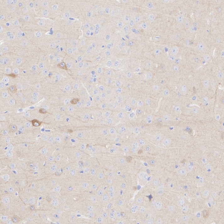
Fig5:
Immunohistochemical analysis of paraffin-embedded mouse cerebral cortex tissue with Rabbit anti-Glutamate receptor 1 antibody (ET1612-10) at 1/1,000 dilution. The section was pre-treated using heat mediated antigen retrieval with Tris-EDTA buffer (pH 9.0) for 20 minutes. The tissues were blocked in 1% BSA for 20 minutes at room temperature, washed with ddH2O and PBS, and then probed with the primary antibody (ET1612-10) at 1/1,000 dilution for 1 hour at room temperature. The detection was performed using an HRP conjugated compact polymer system. DAB was used as the chromogen. Tissues were counterstained with hematoxylin and mounted with DPX. |

|
Fig6:
Immunohistochemical analysis of paraffin-embedded rat cerebral cortex tissue with Rabbit anti-Glutamate receptor 1 antibody (ET1612-10) at 1/1,000 dilution. The section was pre-treated using heat mediated antigen retrieval with Tris-EDTA buffer (pH 9.0) for 20 minutes. The tissues were blocked in 1% BSA for 20 minutes at room temperature, washed with ddH2O and PBS, and then probed with the primary antibody (ET1612-10) at 1/1,000 dilution for 1 hour at room temperature. The detection was performed using an HRP conjugated compact polymer system. DAB was used as the chromogen. Tissues were counterstained with hematoxylin and mounted with DPX. |

|
Fig7: Immunohistochemical analysis of paraffin-embedded mouse cerebellum tissue using anti-Glutamate receptor 1 antibody. The section was pre-treated using heat mediated antigen retrieval with Tris-EDTA buffer (pH 8.0-8.4) for 20 minutes.The tissues were blocked in 5% BSA for 30 minutes at room temperature, washed with ddH2O and PBS, and then probed with the primary antibody (ET1612-10, 1/50) for 30 minutes at room temperature. The detection was performed using an HRP conjugated compact polymer system. DAB was used as the chromogen. Tissues were counterstained with hematoxylin and mounted with DPX. |
Note: All products are “FOR RESEARCH USE ONLY AND ARE NOT INTENDED FOR DIAGNOSTIC OR THERAPEUTIC USE”.