CD59 Recombinant Rabbit Monoclonal Antibody [JM10-71]

cat.: ET1703-28
| Product Type: | Recombinant Rabbit monoclonal IgG, primary antibodies |
|---|---|
| Species reactivity: | Human |
| Applications: | WB, IP |
| Clonality: | Monoclonal |
| Clone number: | JM10-71 |
| Form: | Liquid |
| Storage condition: | Shipped at 4℃. Store at +4℃ short term (1-2 weeks). It is recommended to aliquot into single-use upon delivery. Store at -20℃ long term. |
| Storage buffer: | 1*TBS (pH7.4), 0.05% BSA, 40% Glycerol. Preservative: 0.05% Sodium Azide. |
| Concentration: | 1ug/ul |
| Purification: | Protein A affinity purified. |
| Molecular weight: | Predicted band size: 14 kDa |
| Isotype: | IgG |
| Immunogen: | Synthetic peptide within Human CD59 aa 85-121 / 128. |
| Positive control: | Human placenta tissue lysate, HUVEC cell lysate, K562 cell lysate. |
| Subcellular location: | Cell membrane. Secreted. |
| Recommended Dilutions:
WB IP |
1:1,000-1:5,000 1:10-1:50 |
| Uniprot #: | SwissProt: P13987 Human | O55186 Mouse |
| Alternative names: | 16.3A5 1F5 1F5 antigen 20 kDa homologous restriction factor CD 59 CD_antigen=CD59 CD59 CD59 antigen CD59 antigen complement regulatory protein CD59 antigen p18 20 CD59 antigen p18-20 (antigen identified by monoclonal antibodies 16.3A5, EJ16, EJ30, EL32 and G344) CD59 glycoprotein CD59 molecule CD59 molecule complement regulatory protein CD59_HUMAN Cd59a Complement regulatory protein EJ16 EJ30 EL32 FLJ38134 FLJ92039 G344 HRF 20 HRF-20 HRF20 Human leukocyte antigen MIC11 Ly 6 like protein Lymphocytic antigen CD59/MEM43 MAC inhibitory protein MAC IP MAC-inhibitory protein MAC-IP MACIF MACIP MEM43 MEM43 antigen Membrane attack complex (MAC) inhibition factor Membrane attack complex inhibition factor Membrane inhibitor of reactive lysis MGC2354 MIC11 MIN1 MIN2 MIN3 MIRL MSK21 p18 20 Protectin Surface antigen recognized by monoclonal 16.3A5 T cell activating protein |
Images

|
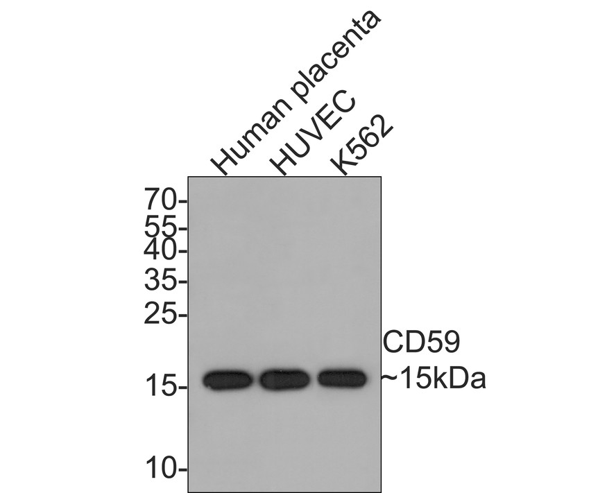
Fig1:
Western blot analysis of CD59 on different lysates with Rabbit anti-CD59 antibody (ET1703-28) at 1/2,000 dilution. Lane 1: Human placenta tissue lysate (20 µg/Lane) Lane 2: HUVEC cell lysate (10 µg/Lane) Lane 3: K562 cell lysate (10 µg/Lane) Predicted band size: 14 kDa Observed band size: 15 kDa Exposure time: 2 minutes; 15% SDS-PAGE gel. Proteins were transferred to a PVDF membrane and blocked with 5% NFDM/TBST for 1 hour at room temperature. The primary antibody (ET1703-28) at 1/2,000 dilution was used in 5% NFDM/TBST at room temperature for 2 hours. Goat Anti-Rabbit IgG - HRP Secondary Antibody (HA1001) at 1:300,000 dilution was used for 1 hour at room temperature. |
Note: All products are “FOR RESEARCH USE ONLY AND ARE NOT INTENDED FOR DIAGNOSTIC OR THERAPEUTIC USE”.