TIMP2 Recombinant Rabbit Monoclonal Antibody [JM87-10]

cat.: ET1703-81
| Product Type: | Recombinant Rabbit monoclonal IgG, primary antibodies |
|---|---|
| Species reactivity: | Human |
| Applications: | WB, FC |
| Clonality: | Monoclonal |
| Clone number: | JM87-10 |
| Form: | Liquid |
| Storage condition: | Store at +4℃ after thawing. Aliquot store at -20℃ or -80℃. Avoid repeated freeze / thaw cycles. |
| Storage buffer: | 1*TBS (pH7.4), 0.05% BSA, 40% Glycerol. Preservative: 0.05% Sodium Azide. |
| Concentration: | 1ug/ul |
| Purification: | Protein A affinity purified. |
| Molecular weight: | Predicted band size: 24 kDa |
| Isotype: | IgG |
| Immunogen: | Synthetic peptide within Human TIMP2 aa 171-220 / 220. |
| Positive control: | K562 cell lysates, A172 cell lysates, Hela. |
| Subcellular location: | Secreted. |
| Recommended Dilutions:
WB FC |
1:500-1:2,000 1:10-1:50 |
| Uniprot #: | SwissProt: P16035 Human |
| Alternative names: | CSC 21K CSC-21K CSC21K Metalloproteinase inhibitor 2 Metalloproteinase inhibitor 2 precursor TIMP 2 TIMP metallopeptidase inhibitor 2 TIMP-2 TIMP2 TIMP2_HUMAN Tissue Inhibitor of Metalloproteinase 2 Tissue inhibitor of metalloproteinases 2 |
Images
|
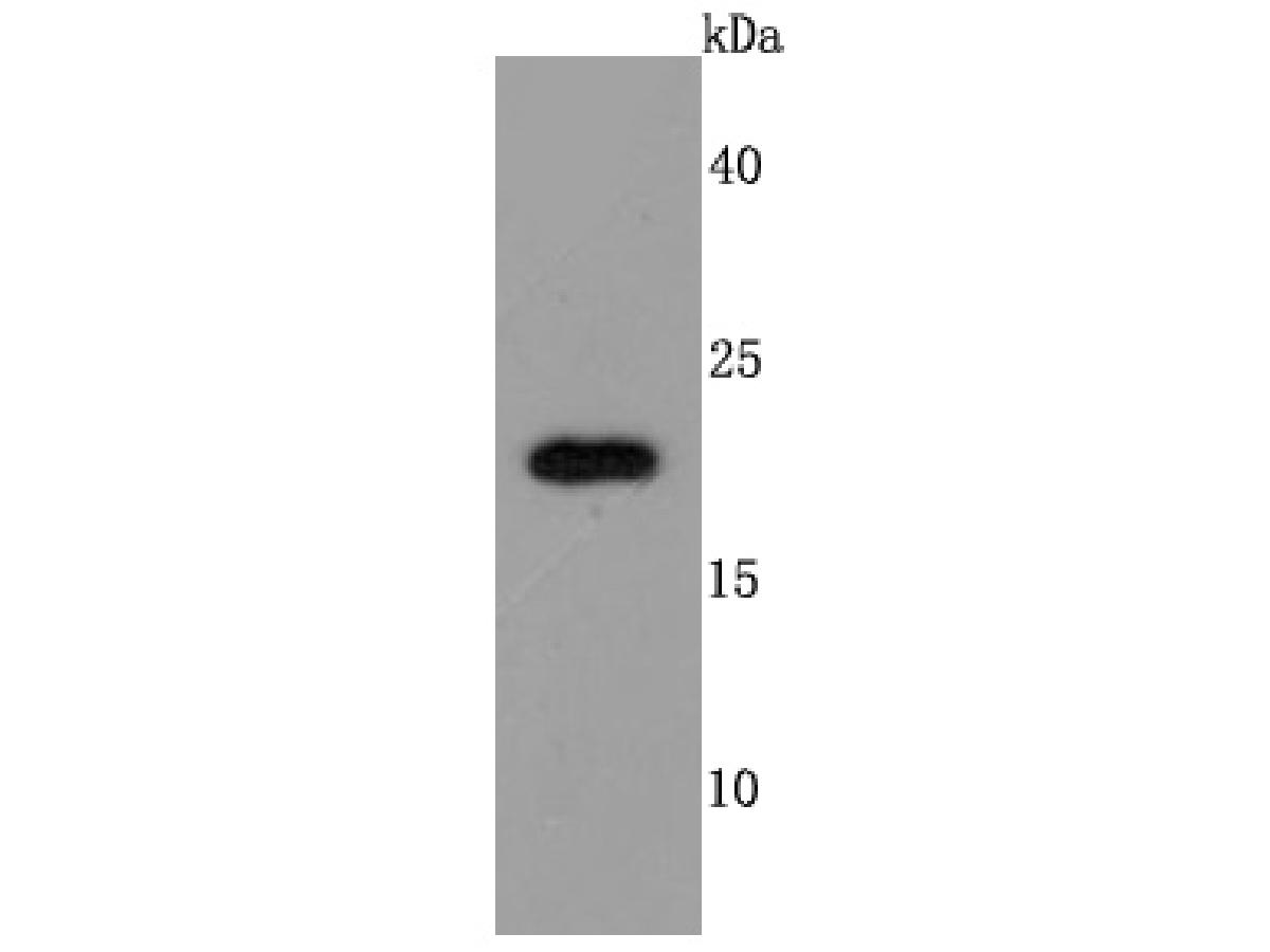
Fig1: Western blot analysis of TIMP2 on K562 cell lysates. Proteins were transferred to a PVDF membrane and blocked with 5% BSA in PBS for 1 hour at room temperature. The primary antibody (ET1703-81, 1/500) was used in 5% BSA at room temperature for 2 hours. Goat Anti-Rabbit IgG - HRP Secondary Antibody (HA1001) at 1:200,000 dilution was used for 1 hour at room temperature. |
|
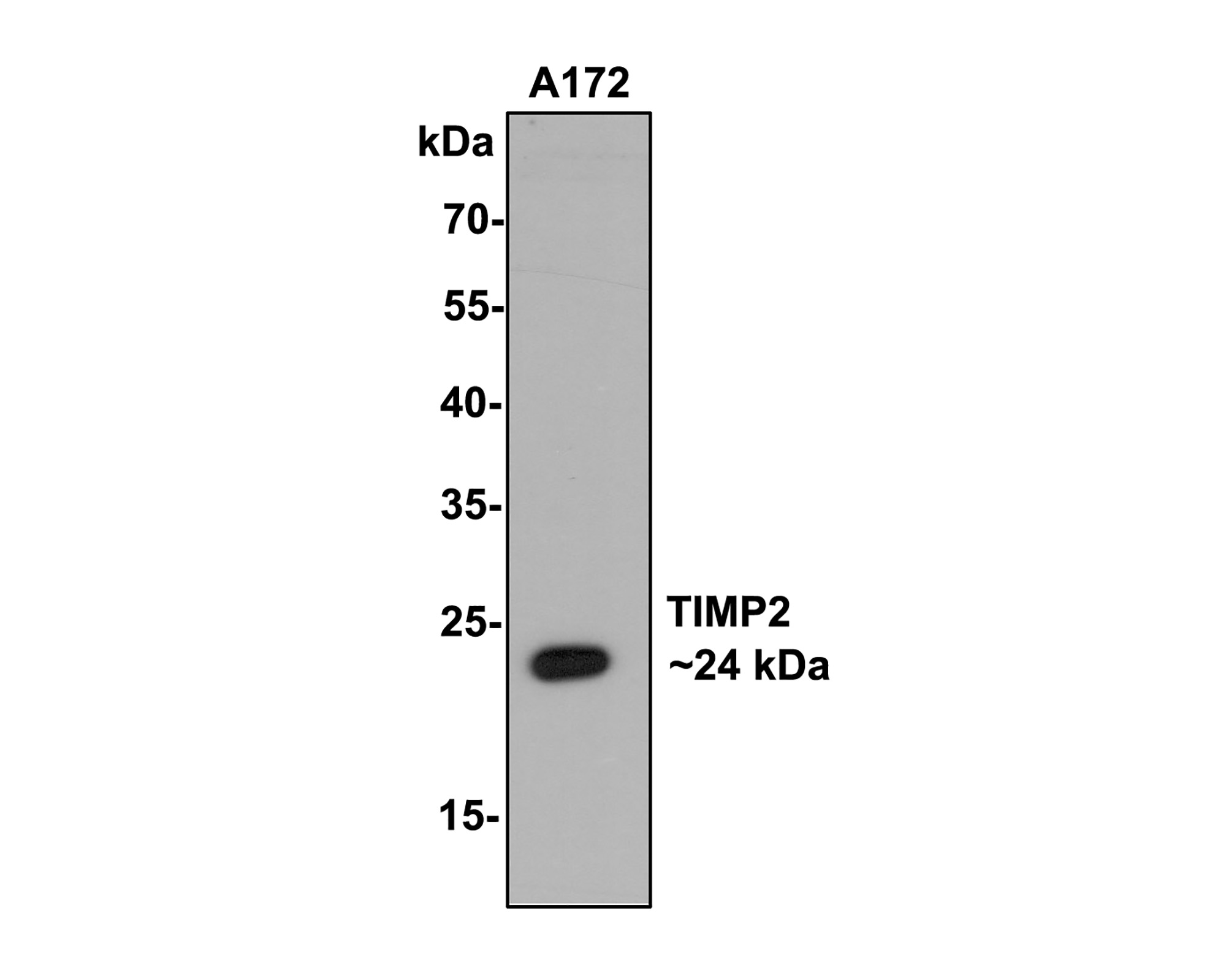
Fig2:
Western blot analysis of TIMP2 on A172 cell lysates with Rabbit anti-TIMP2 antibody (ET1703-81) at 1/500 dilution. Lysates/proteins at 10 µg/Lane. Predicted band size: 24 kDa Observed band size: 24 kDa Exposure time: 2 minutes; 12% SDS-PAGE gel. Proteins were transferred to a PVDF membrane and blocked with 5% NFDM/TBST for 1 hour at room temperature. The primary antibody (ET1703-81) at 1/500 dilution was used in 5% NFDM/TBST at room temperature for 2 hours. Goat Anti-Rabbit IgG - HRP Secondary Antibody (HA1001) at 1:200,000 dilution was used for 1 hour at room temperature. |
|
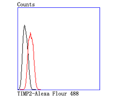
Fig3: Flow cytometric analysis of TIMP2 was done on Hela cells. The cells were fixed, permeabilized and stained with the primary antibody (ET1703-81, 1/50) (red). After incubation of the primary antibody at room temperature for an hour, the cells were stained with a Alexa Fluor 488-conjugated Goat anti-Rabbit IgG Secondary antibody at 1/1000 dilution for 30 minutes.Unlabelled sample was used as a control (cells without incubation with primary antibody; black). |
Note: All products are “FOR RESEARCH USE ONLY AND ARE NOT INTENDED FOR DIAGNOSTIC OR THERAPEUTIC USE”.