CENPB Mouse Monoclonal Antibody [A5F6]

cat.: HA600071
| Product Type: | Mouse monoclonal IgG2a, primary antibodies |
|---|---|
| Species reactivity: | Human, Mouse, Rat |
| Applications: | WB, IF-Cell, IHC-P |
| Clonality: | Monoclonal |
| Clone number: | A5F6 |
| Form: | Liquid |
| Storage condition: | Shipped at 4℃. Store at +4℃ short term (1-2 weeks). It is recommended to aliquot into single-use upon delivery. Store at -20℃ long term. |
| Storage buffer: | 1*TBS (pH7.4), 0.2% BSA, 50% Glycerol. Preservative: 0.05% Sodium Azide. |
| Concentration: | 2ug/ul |
| Purification: | Protein G affinity purified. |
| Molecular weight: | Predicted band size: 65 kDa |
| Isotype: | IgG2a |
| Immunogen: | Recombinant protein within human CENPB aa 1-200 / 599. |
| Positive control: | A431 cell lysate, Daudi cell lysate, 3T3-L1 cell lysate, PC-12 cell lysate, SH-SY5Y, human kidney tissue, human skin tissue. |
| Subcellular location: | Nucleus, centromere. |
| Recommended Dilutions:
WB IF-Cell IHC-P |
1:500-1:2,000 1:50-1:100 1:100-1:500 |
| Uniprot #: | SwissProt: P07199 Human | P27790 Mouse Entrez Gene: 362217 Rat |
| Alternative names: | CENP B CENP-B CENPB CENPB_HUMAN centromere autoantigen B Centromere protein B Centromere protein B, 80kDa Major centromere autoantigen B |
Images
|
Fig1:
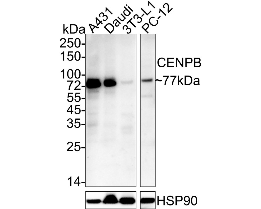
Western blot analysis of CENPB on different lysates with Mouse anti-CENPB antibody (HA600071) at 1/2,000 dilution. Lane 1: A431 cell lysate Lane 2: Daudi cell lysate Lane 3: 3T3-L1 cell lysate Lane 4: PC-12 cell lysate Lysates/proteins at 30 µg/Lane. Predicted band size: 65 kDa Observed band size: 77 kDa Exposure time: 2 minutes; ECL: K1802; 4-20% SDS-PAGE gel. Proteins were transferred to a PVDF membrane and blocked with 5% NFDM/TBST for 1 hour at room temperature. The primary antibody (HA600071) at 1/2,000 dilution was used in 5% NFDM/TBST at 4℃ overnight. Goat Anti-Mouse IgG - HRP Secondary Antibody (HA1006) at 1/50,000 dilution was used for 1 hour at room temperature. |
|
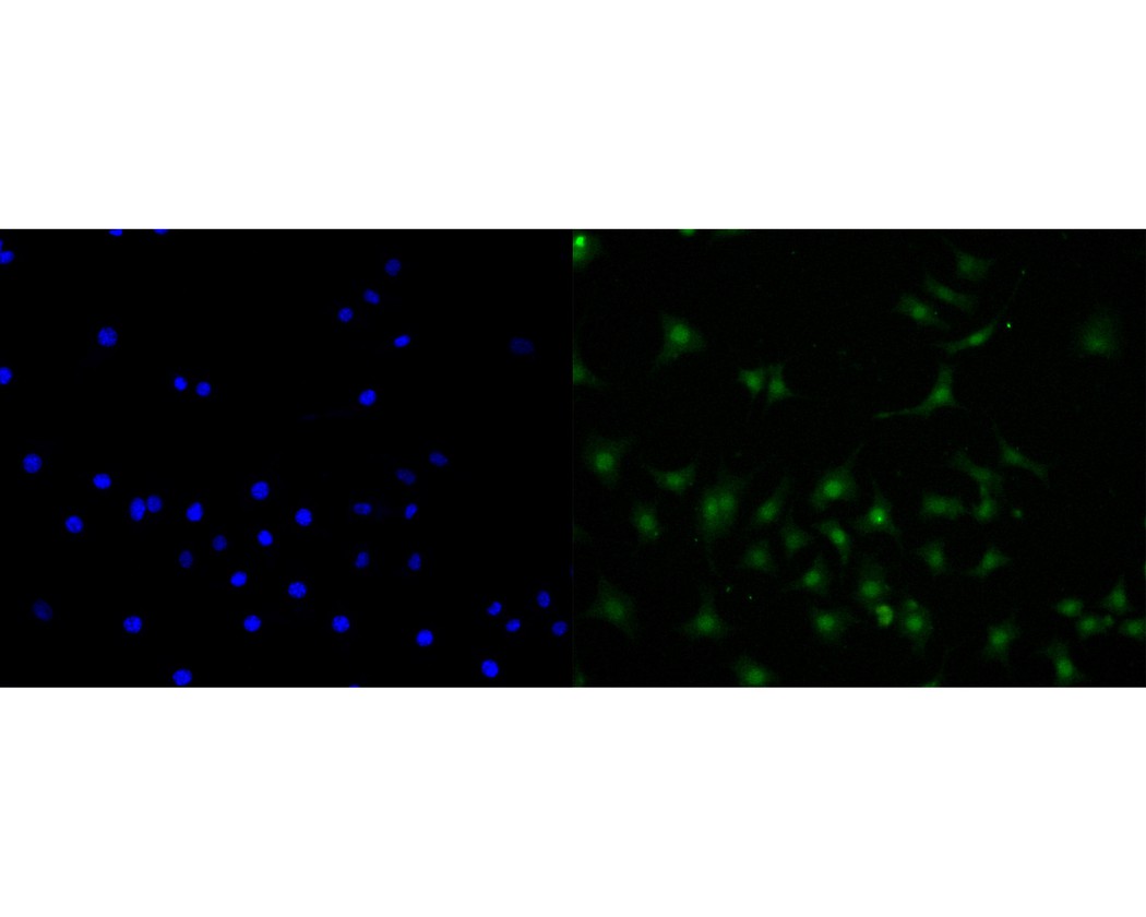
Fig2: ICC staining of CENPB in SH-SY5Y cells (green). Methanol fixed cells were blocked with 10% negative goat serum for 15 minutes at room temperature. Cells were probed with the primary antibody (HA600071, 1/50) for 1 hour at room temperature, washed with PBS. Alexa Fluor®488 conjugate-Goat anti-Mouse IgG was used as the secondary antibody at 1/1,000 dilution. The nuclear counter stain is DAPI (blue). |
|
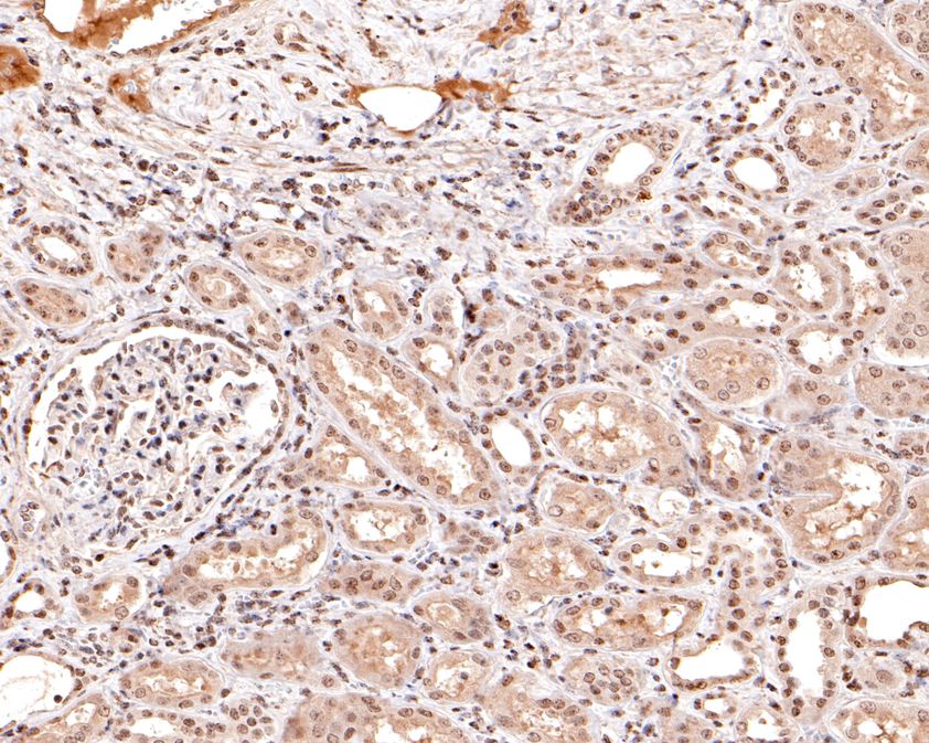
Fig3: Immunohistochemical analysis of paraffin-embedded human kidney tissue using anti-CENPB antibody. The section was pre-treated using heat mediated antigen retrieval with sodium citrate buffer (pH 6.0) for 20 minutes. The tissues were blocked in 1% BSA for 30 minutes at room temperature, washed with ddH2O and PBS, and then probed with the primary antibody (HA600071, 1/400) for 30 minutes at room temperature. The detection was performed using an HRP conjugated compact polymer system. DAB was used as the chromogen. Tissues were counterstained with hematoxylin and mounted with DPX. |
|
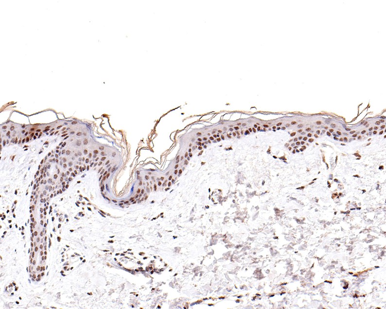
Fig4: Immunohistochemical analysis of paraffin-embedded human skin tissue using anti-CENPB antibody. The section was pre-treated using heat mediated antigen retrieval with sodium citrate buffer (pH 6.0) for 20 minutes. The tissues were blocked in 1% BSA for 30 minutes at room temperature, washed with ddH2O and PBS, and then probed with the primary antibody (HA600071, 1/100) for 30 minutes at room temperature. The detection was performed using an HRP conjugated compact polymer system. DAB was used as the chromogen. Tissues were counterstained with hematoxylin and mounted with DPX. |
Note: All products are “FOR RESEARCH USE ONLY AND ARE NOT INTENDED FOR DIAGNOSTIC OR THERAPEUTIC USE”.