BSA Mouse Monoclonal Antibody [A7D10]

cat.: HA600085
| Product Type: | Mouse monoclonal IgG1, primary antibodies |
|---|---|
| Species reactivity: | Cow |
| Applications: | ELISA, WB |
| Clonality: | Monoclonal |
| Clone number: | A7D10 |
| Form: | Liquid |
| Storage condition: | Store at +4℃ after thawing. Aliquot store at -20℃. Avoid repeated freeze / thaw cycles. |
| Storage buffer: | PBS (pH7.4). |
| Concentration: | 2ug/ul |
| Purification: | Protein G affinity purified. |
| Molecular weight: | Predicted band size: 66 kDa |
| Isotype: | IgG1 |
| Immunogen: | Native protein. |
| Positive control: | BSA protein. |
| Subcellular location: | Secreted. |
| Recommended Dilutions:
ELISA WB |
1:10,000 1:500 |
| Uniprot #: | SwissProt: P02769 Bovine |
| Alternative names: | Albumin BSA Bos d 6 ALB |
Images
|
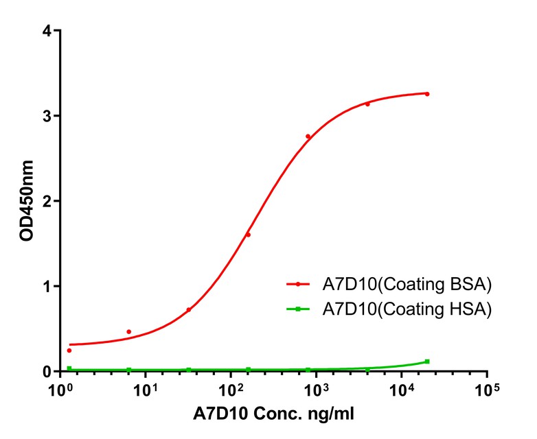
Fig1:
Bovine Serum Albumin Antibody (HA600085) in indirect ELISA. Indirect ELISA analysis of BSA was performed by coating wells of a 96-well plate with 100 µl per well of BSA standard diluted in carbonate/bicarbonate buffer, at a concentration of 1 µg/mL overnight at 4℃. Wells of the plate were washed, blocked with StartingBlock blocking buffer, and incubated with 100 µl per well of a mouse BSA monoclonal antibody starting at a concentration of 20 µg/mL and serially diluting it to a concentration of 1.28 ng/mL for 2 hours at room temperature. The plate was washed and incubated with 100 µl per well of an HRP-conjugated goat anti-mouse IgG secondary antibody at a dilution of 1:10,000 for one hour at room temperature. Detection was performed using an Ultra TMB Substrate for 5 minutes at room temperature in the dark. The reaction was stopped with sulfuric acid and absorbances were read on a spectrophotometer at 450 nm. |
|
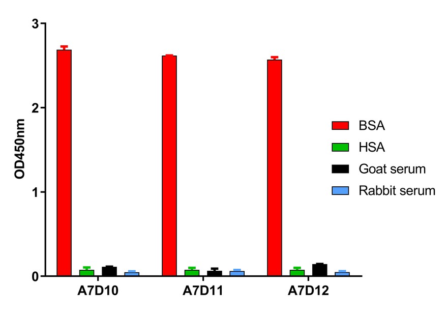
Fig2:
Bovine Serum Albumin Antibody (HA600085) in indirect ELISA. Indirect ELISA analysis of BSA was performed by coating wells of a 96-well plate with 100 µl per well of BSA standard diluted in carbonate/bicarbonate buffer, at a concentration of 1 µg/mL overnight at 4℃. Wells of the plate were washed, blocked with StartingBlock blocking buffer, and incubated with 100 µl per well of a mouse BSA monoclonal antibody starting at a concentration of 0.8 µg/mL for 2 hours at room temperature. The plate was washed and incubated with 100 µl per well of an HRP-conjugated goat anti-mouse IgG secondary antibody at a dilution of 1:10,000 for one hour at room temperature. Detection was performed using an Ultra TMB Substrate for 5 minutes at room temperature in the dark. The reaction was stopped with sulfuric acid and absorbances were read on a spectrophotometer at 450 nm. This antibody demonstrates high specificity for BSA and little or no crossreactivity to HSA (Human serum albumin), Goat serum and Rabbit serum. |
|
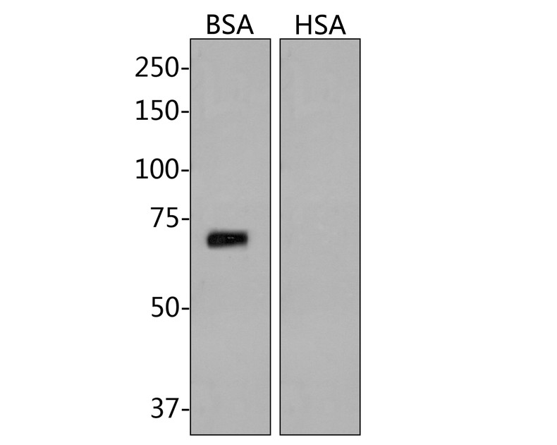
Fig3:
Western blot analysis of BSA on BSA protein with Mouse anti-BSA antibody (HA600085) at 1/500 dilution. Lysates/proteins at 50 µg/Lane. Predicted band size: 66 kDa Observed band size: 70 kDa Exposure time: 2 minutes; 8% SDS-PAGE gel. Proteins were transferred to a PVDF membrane and blocked with 5% NFDM/TBST for 1 hour at room temperature. The primary antibody (HA600085) at 1/500 dilution was used in 5% NFDM/TBST at room temperature for 2 hours. Goat Anti-Mouse IgG - HRP Secondary Antibody (HA1006) at 1:100,000 dilution was used for 1 hour at room temperature. |
Note: All products are “FOR RESEARCH USE ONLY AND ARE NOT INTENDED FOR DIAGNOSTIC OR THERAPEUTIC USE”.