iFluor™ 647 Conjugated MUC1 Mouse Monoclonal Antibody [PSH0-49]

cat.: HA600108F
| Product Type: | Mouse monoclonal IgG2a, primary antibodies |
|---|---|
| Species reactivity: | Human |
| Applications: | FC |
| Clonality: | Monoclonal |
| Clone number: | PSH0-49 |
| Form: | Liquid |
| Storage condition: | Shipped at 4℃. Store at +4℃ short term (1-2 weeks). It is recommended to aliquot into single-use upon delivery. Store at -20℃ long term. |
| Storage buffer: | Preservative: 0.02% Sodium azide Constituents: 30% Glycerol, 1% BSA, 68.98% PBS |
| Concentration: | 1ug/ul |
| Purification: | Protein A affinity purified. |
| Molecular weight: | Predicted band size: 122 kDa |
| Isotype: | IgG2a |
| Immunogen: | Synthetic peptide of core peptide domain of human MUC1. |
| Positive control: | MCF7, SK-Br-3. |
| Subcellular location: | Apical cell membrane. Secreted. Nucleus, Cell membrane, Cytoplasm. |
| Recommended Dilutions:
FC |
1:500-1:1,000 |
| Uniprot #: | SwissProt: P15941 Human |
| Alternative names: | ADMCKD ADMCKD1 Breast carcinoma associated antigen DF3 Breast carcinoma-associated antigen DF3 CA 15-3 CA15 3 CA15 3 antigen CA15-3 CA15.3 Cancer antigen 15-3 Carcinoma associated mucin Carcinoma-associated mucin CD 227 CD227 DF3 antigen EMA Episialin Epithelial Membrane Antigen H23 antigen H23AG KL 6 KL-6 KL6 Krebs von den Lungen-6 MAM 6 MAM6 MCD MCKD MCKD1 Medullary cystic kidney disease 1 (autosomal dominant) Medullary cystic kidney disease, autosomal dominant MUC 1 MUC-1 MUC-1/SEC MUC-1/X MUC1 MUC1-alpha MUC1-beta MUC1-CT MUC1-NT MUC1/ZD MUC1_HUMAN Mucin 1 Mucin 1 cell surface associated Mucin 1 transmembrane Mucin 1, cell surface associated Mucin-1 subunit beta Peanut reactive urinary mucin Peanut-reactive urinary mucin PEM PEMT Polymorphic epithelial mucin PUM Tumor associated epithelial membrane antigen Tumor associated epithelial mucin Tumor associated mucin Tumor-ass...... |
Images
|
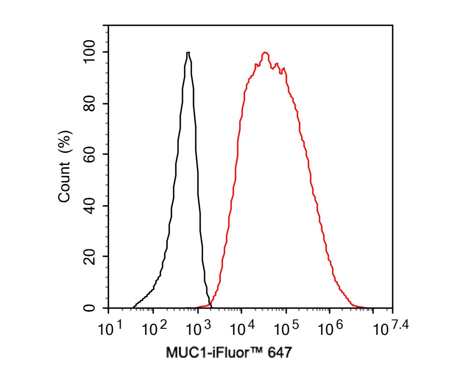
Fig1:
Flow cytometric analysis of MCF7 cells labeling MUC1. Cells were washed twice with cold PBS and resuspend. Then incubated for 1 hour at +4℃ with MUC1 (HA600108F, red, 1ug/ml). Unlabelled sample was used as a control (cells without incubation with primary antibody; black). |
|
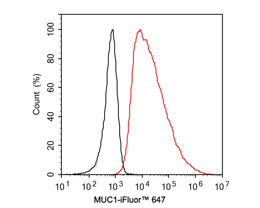
Fig2:
Flow cytometric analysis of SK-Br-3 cells labeling MUC1. Cells were washed twice with cold PBS and resuspend. Then incubated for 1 hour at +4℃ with MUC1 (HA600108F, red, 1ug/ml). Unlabelled sample was used as a control (cells without incubation with primary antibody; black). |
Note: All products are “FOR RESEARCH USE ONLY AND ARE NOT INTENDED FOR DIAGNOSTIC OR THERAPEUTIC USE”.