iFluor™ 488 Conjugated E-Cadherin Recombinant Rabbit Monoclonal Antibody [SY0287]

cat.: HA720159F
| Product Type: | Recombinant Rabbit monoclonal IgG, primary antibodies |
|---|---|
| Species reactivity: | Human |
| Applications: | IF-Cell, FC |
| Clonality: | Monoclonal |
| Clone number: | SY0287 |
| Form: | Liquid |
| Storage condition: | Shipped at 4℃. Store at +4℃ short term (1-2 weeks). It is recommended to aliquot into single-use upon delivery. Store at -20℃ long term. |
| Storage buffer: | Preservative: 0.02% Sodium azide Constituents: 30% Glycerol, 1% BSA, 68.98% PBS |
| Concentration: | 1ug/ul |
| Purification: | Protein A affinity purified. |
| Molecular weight: | Predicted band size: 97 kDa |
| Isotype: | IgG |
| Immunogen: | Synthetic peptide within Human E-Cadherin aa 591-640 / 882. |
| Positive control: | MCF-7, A431. |
| Subcellular location: | Endosome, Cell membrane, trans-Golgi network, adherens junction. |
| Recommended Dilutions:
IF-Cell FC |
1:100 1:50-1:1,000 |
| Uniprot #: | SwissProt: P12830 Human |
| Alternative names: | Arc 1 CADH1_HUMAN Cadherin 1 cadherin 1 type 1 E-cadherin Cadherin1 CAM 120/80 CD 324 CD324 CD324 antigen cdh1 CDHE E-Cad/CTF3 E Cadherin E-cadherin ECAD Epithelial cadherin epithelial calcium dependant adhesion protein LCAM Liver cell adhesion molecule UVO Uvomorulin |
Images
|
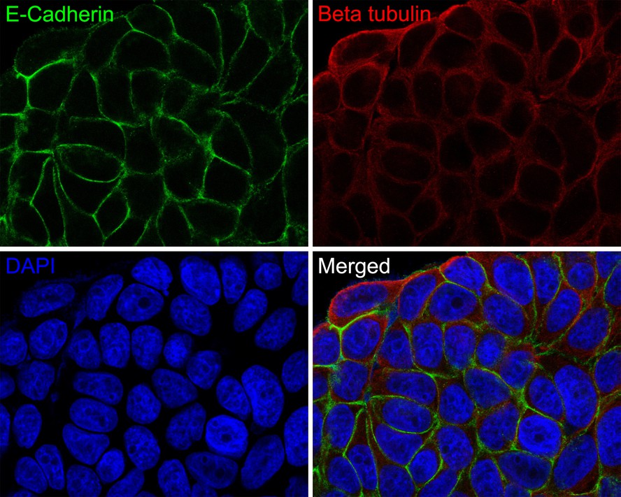
Fig1:
Immunocytochemistry analysis of MCF-7 cells labeling E-Cadherin with Rabbit anti-E-Cadherin antibody (HA720159F) at 1/100 dilution. Cells were fixed in 4% paraformaldehyde for 10 minutes, permeabilized with 0.1% Triton X-100 in PBS for 15 minutes, and then blocked with 2% normal goat serum for 1 hour at 37 ℃. Cells were then incubated with Rabbit anti-E-Cadherin antibody (HA720159F) at 1/100 dilution in 2% normal goat serum overnight at 4 ℃. Nuclear DNA was labelled in blue with DAPI. Beta tubulin (M1305-2, red) was stained at 1/200 dilution overnight at +4℃. Goat Anti-Mouse IgG H&L (iFluor™ 594, HA1126) were used as the secondary antibody at 1/800 dilution. |
|
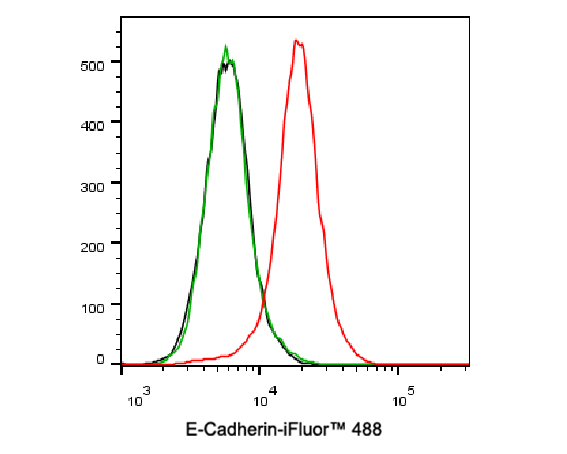
Fig2:
Flow cytometric analysis of A431 cells labeling E-Cadherin. Cells were washed twice with cold PBS and resuspend. Then incubated for 30 minutes at +4℃ with E-Cadherin (HA720159F, red, 1ug/ml) and Rabbit IgG Isotype Control (iFluor™ 488, green, 1ug/ml). Unlabelled sample was used as a control (cells without incubation with primary antibody; black). |
|
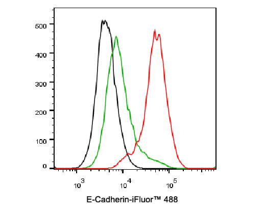
Fig3:
Flow cytometric analysis of MCF-7 cells labeling E-Cadherin. Cells were washed twice with cold PBS and resuspend. Then incubated for 30 minutes at +4℃ with E-Cadherin (HA720159F, red, 10ug/ml) and Rabbit IgG Isotype Control (iFluor™ 488, green, 10ug/ml). Unlabelled sample was used as a control (cells without incubation with primary antibody; black). |
Note: All products are “FOR RESEARCH USE ONLY AND ARE NOT INTENDED FOR DIAGNOSTIC OR THERAPEUTIC USE”.