iFluor™ 488 Conjugated Ki67 Recombinant Rabbit Monoclonal Antibody [SR00-02]

cat.: HA720162F
| Product Type: | Recombinant Rabbit monoclonal IgG, primary antibodies |
|---|---|
| Species reactivity: | Human |
| Applications: | IF-Cell, IF-Tissue, FC |
| Clonality: | Monoclonal |
| Clone number: | SR00-02 |
| Form: | Liquid |
| Storage condition: | Shipped at 4℃. Store at +4℃ short term (1-2 weeks). It is recommended to aliquot into single-use upon delivery. Store at -20℃ long term. |
| Storage buffer: | Preservative: 0.02% Sodium azide Constituents: 30% Glycerol, 1% BSA, 68.98% PBS |
| Concentration: | 1ug/ul |
| Purification: | Protein A affinity purified. |
| Molecular weight: | Predicted band size: 359 kDa |
| Isotype: | IgG |
| Immunogen: | Synthetic peptide within human Ki67 aa 1,040-1,080. |
| Positive control: | A431, Jurkat cells, human colon carcinoma tissue, human lymph nodes tissue, Hela. |
| Subcellular location: | Nucleus, Chromosome. |
| Recommended Dilutions:
IF-Cell IF-Tissue FC |
1:100 1:100 1:100-1:1,000 |
| Uniprot #: | SwissProt: P46013 Human |
| Alternative names: | Antigen identified by monoclonal Ki 67 Antigen identified by monoclonal Ki-67 Antigen KI-67 Antigen KI67 Antigen Ki67 KI67_HUMAN KIA Marker of proliferation Ki-67 MIB 1 MIB MKI67 PPP1R105 Proliferation marker protein Ki-67 Proliferation related Ki 67 antigen Protein phosphatase 1 regulatory subunit 105 RP11-380J17.2 |
Images
|
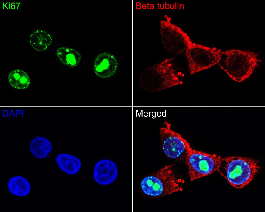
Fig1:
Immunocytochemistry analysis of A431 cells labeling Ki67 with Rabbit anti-Ki67 antibody (HA720162F) at 1/100 dilution. Cells were fixed in 100% methanol for 10 minutes, permeabilized with 0.1% Triton X-100 in PBS for 15 minutes, and then blocked with 1% BSA for 30 minutes at room temperature. Cells were then incubated with Rabbit anti-Ki67 antibody (HA720162F) at 1/100 dilution in 1% BSA overnight at 4 ℃. Nuclear DNA was labelled in blue with DAPI. Beta tubulin (M1305-2, red) was stained at 1/200 dilution overnight at +4℃. Goat Anti-Mouse IgG H&L (iFluor™ 594, HA1126) were used as the secondary antibody at 1/1,000 dilution. |
|
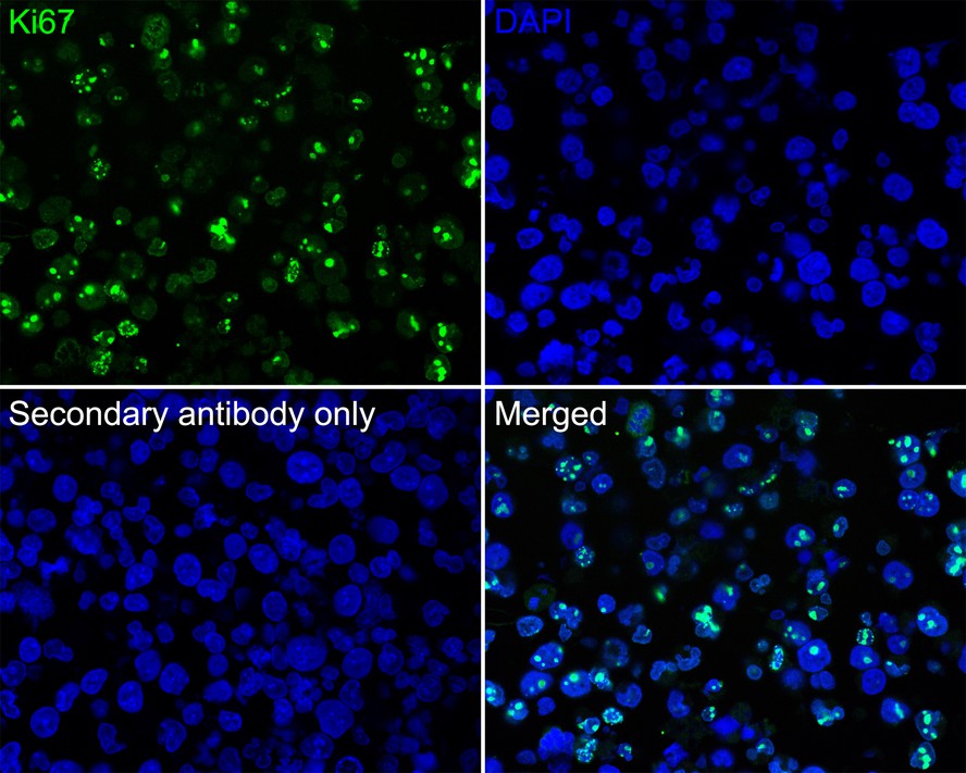
Fig2:
Immunofluorescence analysis of paraffin-embedded Jurkat cells labeling Ki67 with Rabbit anti-Ki67 antibody (HA720162F) at 1/100 dilution. The section was pre-treated using heat mediated antigen retrieval with Tris-EDTA buffer (pH 9.0) for 20 minutes. The tissues were blocked in 10% negative goat serum for 1 hour at room temperature, washed with PBS, and then probed with the primary antibody (HA720162F, green) at 1/100 dilution overnight at 4 ℃, washed with PBS. |
|
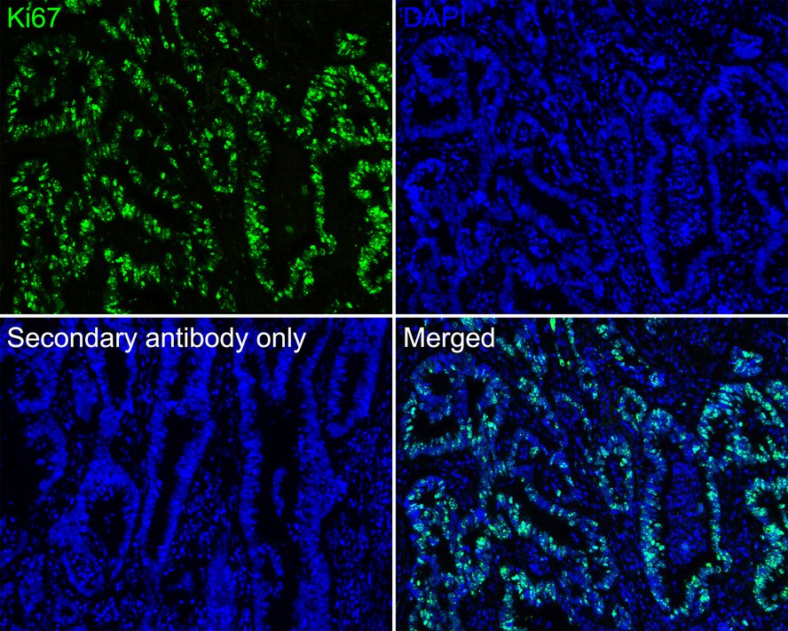
Fig3:
Immunofluorescence analysis of paraffin-embedded human colon carcinoma tissue labeling Ki67 (HA720162F). The section was pre-treated using heat mediated antigen retrieval with sodium citrate buffer (pH 6.0) (high pressure) for 2 minutes. The tissues were blocked in 10% negative goat serum for 1 hour at room temperature, washed with PBS. And then probed with the primary antibody Ki67 (HA720162F, iFluor™ 488) at 1/100 dilution overnight at 4 ℃, washed with PBS. DAPI was used as nuclear counterstain. |
|
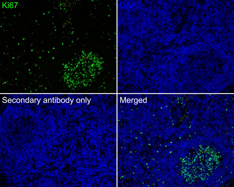
Fig4:
Immunofluorescence analysis of paraffin-embedded human lymph nodes tissue labeling Ki67 (HA720162F). The section was pre-treated using heat mediated antigen retrieval with sodium citrate buffer (pH 6.0) (high pressure) for 2 minutes. The tissues were blocked in 10% negative goat serum for 1 hour at room temperature, washed with PBS. And then probed with the primary antibody Ki67 (HA720162F, iFluor™ 488) at 1/100 dilution overnight at 4 ℃, washed with PBS. DAPI was used as nuclear counterstain. |
|
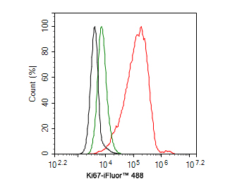
Fig5:
Flow cytometric analysis of A431 cells labeling Ki67. Cells were fixed and permeabilized. Then incubated for 1 hour at +4℃ with Ki67 (HA720162F, red, 1ug/ml) and Rabbit IgG Isotype Control (iFluor™ 488, green, 1ug/ml). Unlabelled sample was used as a control (cells without incubation with primary antibody; black). |
|
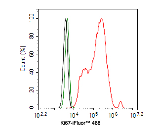
Fig6:
Flow cytometric analysis of Hela cells labeling Ki67. Cells were fixed and permeabilized. Then incubated for 1 hour at +4℃ with Ki67 (HA720162F, red, 10ug/ml) and Rabbit IgG Isotype Control (iFluor™ 488, green, 10ug/ml). Unlabelled sample was used as a control (cells without incubation with primary antibody; black). |
Note: All products are “FOR RESEARCH USE ONLY AND ARE NOT INTENDED FOR DIAGNOSTIC OR THERAPEUTIC USE”.