CCDC47 Mouse Monoclonal Antibody [1-B9-5]

cat.: M1004-3
| Product Type: | Mouse monoclonal IgG1, primary antibodies |
|---|---|
| Species reactivity: | Human, Mouse |
| Applications: | IF-Cell, IHC-P, WB |
| Clonality: | Monoclonal |
| Clone number: | 1-B9-5 |
| Form: | Liquid |
| Storage condition: | Store at +4℃ after thawing. Aliquot store at -20℃ or -80℃. Avoid repeated freeze / thaw cycles. |
| Storage buffer: | 1*PBS (pH7.4), 0.2% BSA, 40% Glycerol. Preservative: 0.05% Sodium Azide. |
| Concentration: | 2ug/ul |
| Purification: | Protein G affinity purified. |
| Molecular weight: | Predicted band size: 56 kDa |
| Isotype: | IgG1 |
| Immunogen: | Recombinant protein within Human CCDC47 aa 1-483 / 483. |
| Positive control: | Recombinant protein, F9, MCF-7, human liver tissue, human lung tissue, human testis tissue, human breast tissue, human colon tissue, human stomach tissue. |
| Subcellular location: | Endoplasmic reticulum membrane. |
| Recommended Dilutions:
WB IF-Cell IHC-P |
1:500-1:1,000 1:50-1:200 1:50-1:200 |
| Uniprot #: | SwissProt: Q96A33 Human | Q9D024 Mouse |
| Alternative names: | CCD47_HUMAN CCDC 47 CCDC47 Coiled coil domain containing 47 Coiled coil domain containing protein 47 Coiled-coil domain-containing protein 47 GK001 MSTP041 |
Images
|
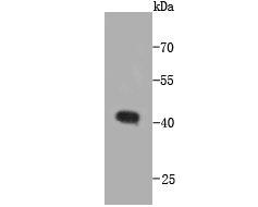
Fig1: Western blot analysis of CCDC47 on recombinant protein using anti-CCDC47 antibody at 1/1,000 dilution. |
|
Fig2:
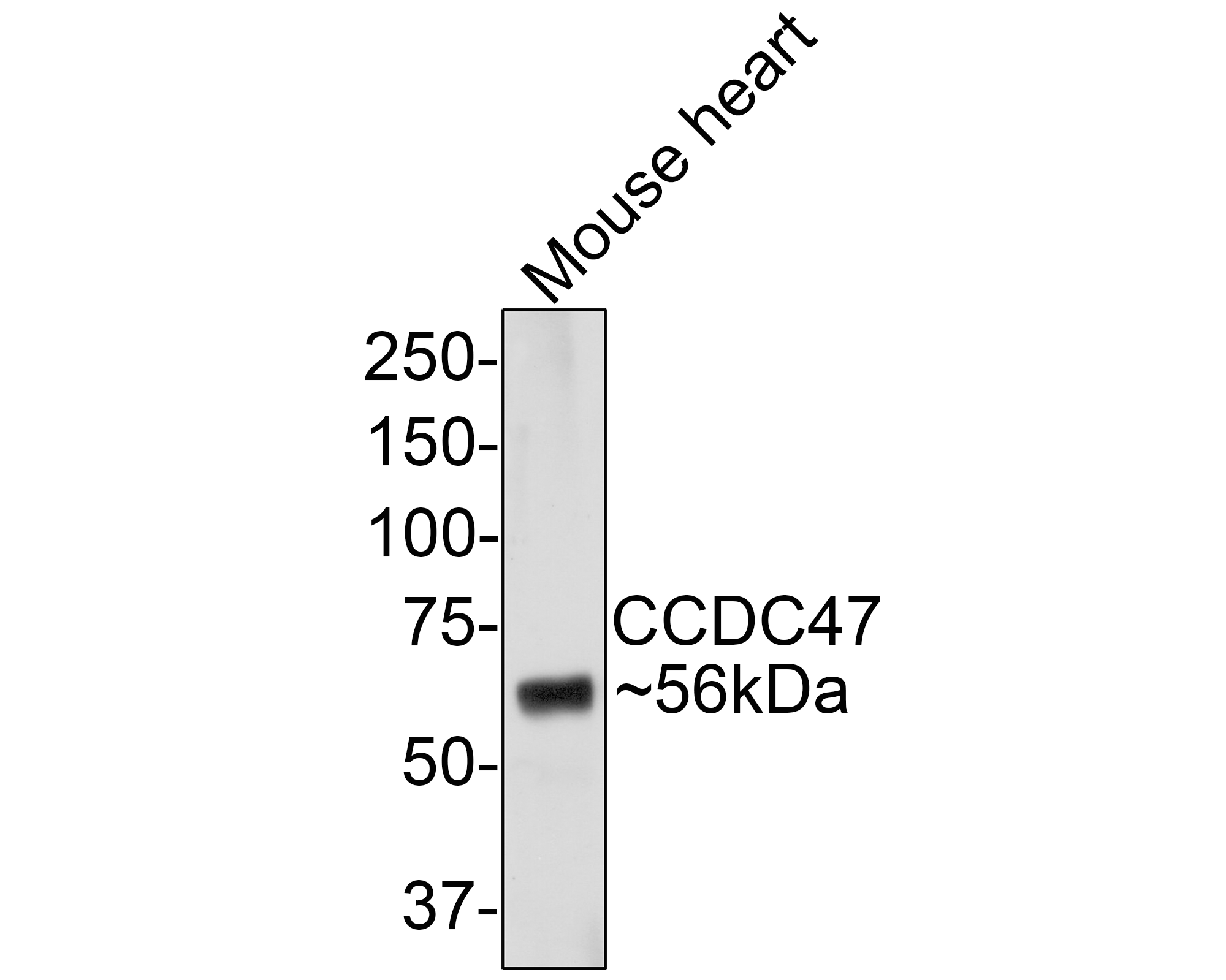
Western blot analysis of CCDC47 on Mouse heart tissue lysates with Mouse anti-CCDC47 antibody (M1004-3) at 1/1,000 dilution. Lysates/proteins at 20 µg/Lane. Predicted band size: 56 kDa Observed band size: 56 kDa Exposure time: 2 minutes; 10% SDS-PAGE gel. Proteins were transferred to a PVDF membrane and blocked with 5% NFDM/TBST for 1 hour at room temperature. The primary antibody (M1004-3) at 1/1,000 dilution was used in 5% NFDM/TBST at room temperature for 2 hours. Goat Anti-Mouse IgG - HRP Secondary Antibody (HA1006) at 1:100,000 dilution was used for 1 hour at room temperature. |
|
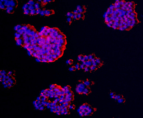
Fig3: ICC staining CCDC47 in F9 cells (red). The nuclear counter stain is DAPI (blue). Cells were fixed in paraformaldehyde, permeabilised with 0.25% Triton X100/PBS. |
|
Fig4:
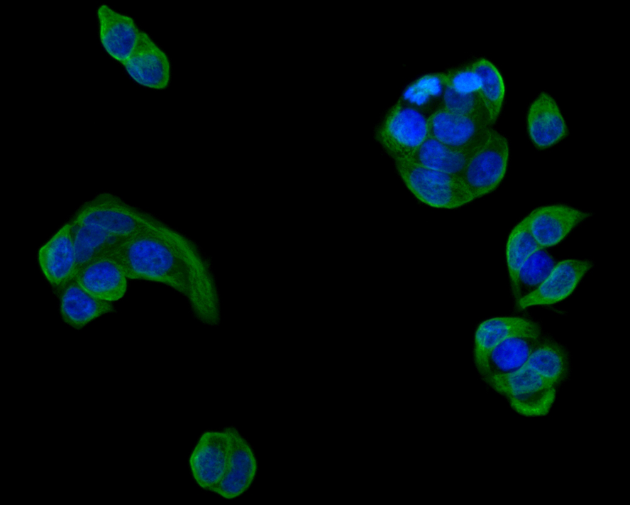
Immunocytochemistry analysis of MCF-7 cells labeling CCDC47 with Mouse anti-CCDC47 antibody (M1004-3) at 1/50 dilution. Cells were fixed in 4% paraformaldehyde for 30 minutes, permeabilized with 0.1% Triton X-100 in PBS for 15 minutes, and then blocked with 2% BSA for 30 minutes at room temperature. Cells were then incubated with Mouse anti-CCDC47 antibody (M1004-3) at 1/50 dilution in 2% BSA overnight at 4 ℃. Goat Anti-Mouse IgG H&L (iFluor™ 488, HA1125) was used as the secondary antibody at 1/1,000 dilution. PBS instead of the primary antibody was used as the secondary antibody only control. Nuclear DNA was labelled in blue with DAPI. |
|
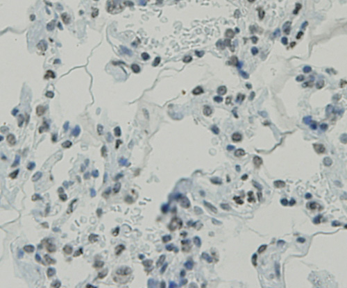
Fig5: Immunohistochemical analysis of paraffin-embedded human lung tissue using anti-CCDC47 antibody. Counter stained with hematoxylin. |
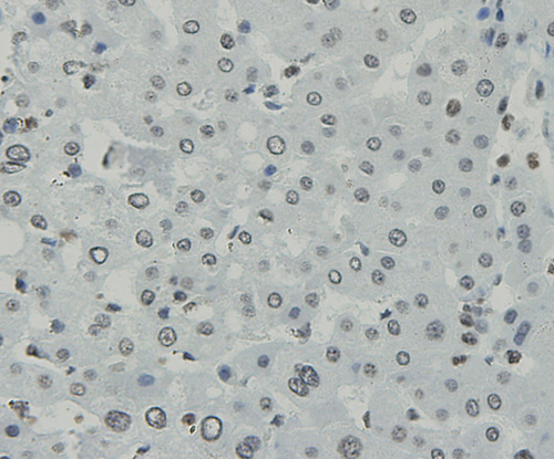
|
Fig6: Immunohistochemical analysis of paraffin-embedded human liver tissue using anti-CCDC47 antibody. Counter stained with hematoxylin. |
|
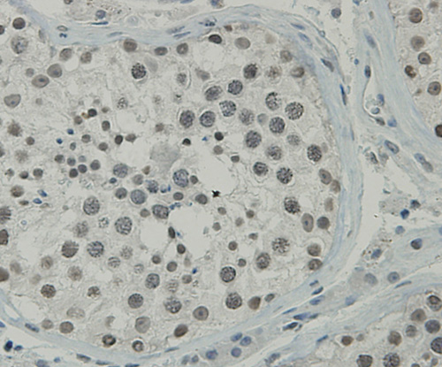
Fig7: Immunohistochemical analysis of paraffin-embedded human testis tissue using anti-CCDC47 antibody. Counter stained with hematoxylin. |
|
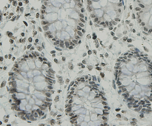
Fig8: Immunohistochemical analysis of paraffin-embedded human colon tissue using anti-CCDC47 antibody. Counter stained with hematoxylin. |
|
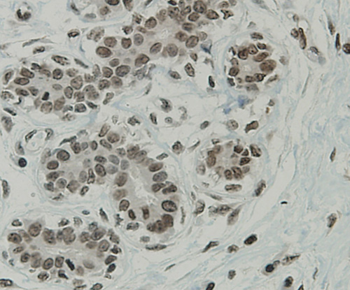
Fig9: Immunohistochemical analysis of paraffin-embedded human breast tissue using anti-CCDC47 antibody. Counter stained with hematoxylin. |
|
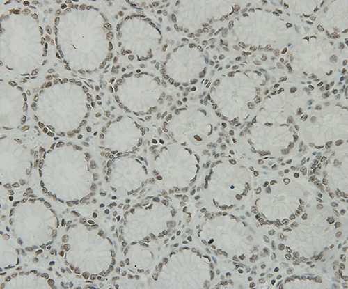
Fig10: Immunohistochemical analysis of paraffin-embedded human stomach tissue using anti-CCDC47 antibody. Counter stained with hematoxylin. |
Note: All products are “FOR RESEARCH USE ONLY AND ARE NOT INTENDED FOR DIAGNOSTIC OR THERAPEUTIC USE”.