BRCA2 Mouse Monoclonal Antibody [A2-B10]

cat.: M1310-4
| Product Type: | Mouse monoclonal IgG1, primary antibodies |
|---|---|
| Species reactivity: | Human |
| Applications: | IF-Cell, IHC-P |
| Clonality: | Monoclonal |
| Clone number: | A2-B10 |
| Form: | Liquid |
| Storage condition: | Store at +4℃ after thawing. Aliquot store at -20℃ or -80℃. Avoid repeated freeze / thaw cycles. |
| Storage buffer: | 1*PBS (pH7.4), 0.2% BSA, 40% Glycerol. Preservative: 0.05% Sodium Azide. |
| Concentration: | 2ug/ul |
| Purification: | Protein A affinity purified. |
| Molecular weight: | 384 kDa |
| Isotype: | IgG1 |
| Immunogen: | Synthetic peptide within Human BRCA2 aa 3,369-3,418 / 3,418. |
| Positive control: | Human breast carcinoma tissue, A549, SKOV-3, MCF-7. |
| Subcellular location: | Nucleus, cytoplasm. |
| Recommended Dilutions:
IF-Cell IHC-P |
1:200 1:150-1:200 |
| Uniprot #: | SwissProt: P51587 Human |
| Alternative names: | BRCA 2 BRCA1/BRCA2 containing complex subunit 2 Brca2 BRCA2, DNA repair associated BRCA2_HUMAN BRCC 2 BRCC2 Breast and ovarian cancer susceptibility gene early onset breast and ovarian cancer susceptibility protein 2 Breast cancer 2 early onset Breast Cancer 2 tumor suppressor Breast cancer susceptibility protein BRCA2 Breast cancer type 2 susceptibility protein BROVCA2 FACD FAD 1 FAD FAD1 FANCB FANCD 1 FANCD FANCD1 FANCD1 gene Fanconi anemia complementation group D1 Fanconi anemia group D1 protein GLM3 mutant BRCA2 OTTHUMP00000018803 OTTHUMP00000042401 PNCA2 XRCC11 |
Images
|
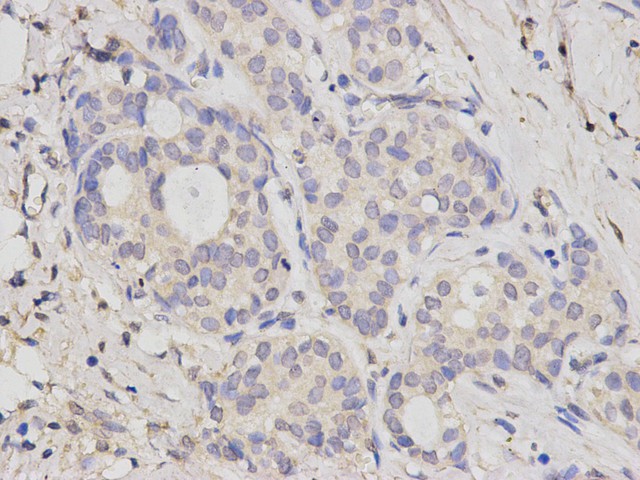
Fig1: Immunohistochemical analysis of paraffin-embedded human breast carcinoma tissue using anti-BRCA2 antibody. The section was pre-treated using heat mediated antigen retrieval with sodium citrate buffer (pH 6.0) for 20 minutes. The tissues were blocked in 5% BSA for 30 minutes at room temperature, washed with ddH2O and PBS, and then probed with the primary antibody (M1310-4, 1/200) for 30 minutes at room temperature. The detection was performed using an HRP conjugated compact polymer system. DAB was used as the chromogen. Tissues were counterstained with hematoxylin and mounted with DPX. |
|
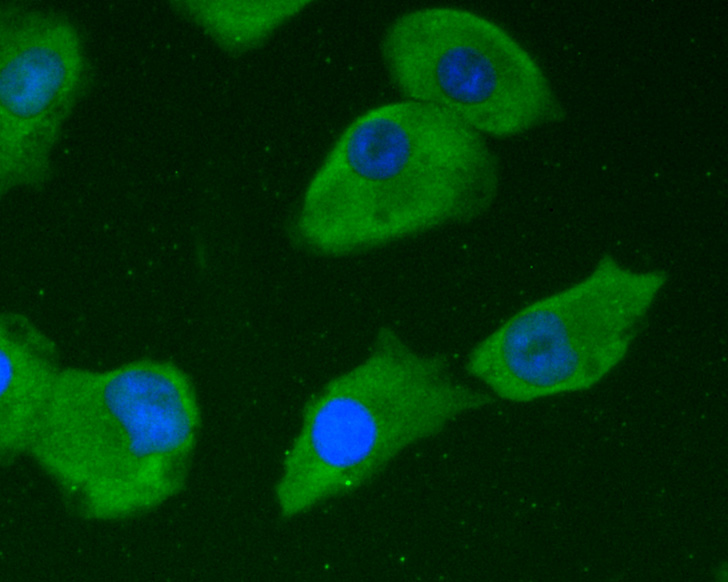
Fig2: ICC staining of BRCA2 in A549 cells (green). Formalin fixed cells were permeabilized with 0.1% Triton X-100 in TBS for 10 minutes at room temperature and blocked with 1% Blocker BSA for 15 minutes at room temperature. Cells were probed with the primary antibody (M1310-4, 1/50) for 1 hour at room temperature, washed with PBS. Alexa Fluor®488 Goat anti-Mouse IgG was used as the secondary antibody at 1/1,000 dilution. The nuclear counter stain is DAPI (blue). |
|
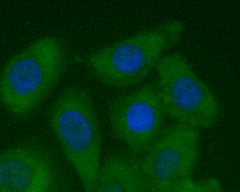
Fig3: ICC staining of BRCA2 in SKOV-3 cells (green). Formalin fixed cells were permeabilized with 0.1% Triton X-100 in TBS for 10 minutes at room temperature and blocked with 1% Blocker BSA for 15 minutes at room temperature. Cells were probed with the primary antibody (M1310-4, 1/50) for 1 hour at room temperature, washed with PBS. Alexa Fluor®488 Goat anti-Mouse IgG was used as the secondary antibody at 1/1,000 dilution. The nuclear counter stain is DAPI (blue). |
|
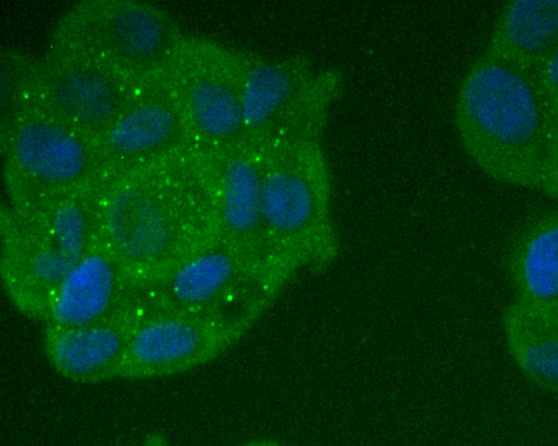
Fig4: ICC staining of BRCA2 in MCF-7 cells (green). Formalin fixed cells were permeabilized with 0.1% Triton X-100 in TBS for 10 minutes at room temperature and blocked with 1% Blocker BSA for 15 minutes at room temperature. Cells were probed with the primary antibody (M1310-4, 1/200) for 1 hour at room temperature, washed with PBS. Alexa Fluor®488 Goat anti-Mouse IgG was used as the secondary antibody at 1/1,000 dilution. The nuclear counter stain is DAPI (blue). |
Note: All products are “FOR RESEARCH USE ONLY AND ARE NOT INTENDED FOR DIAGNOSTIC OR THERAPEUTIC USE”.