Caspase-1 Mouse Monoclonal Antibody [B0-6]

cat.: M1505-2
| Product Type: | Mouse monoclonal IgG1, primary antibodies |
|---|---|
| Species reactivity: | Human, Mouse, Rat |
| Applications: | WB, IF-Cell, IHC-P |
| Clonality: | Monoclonal |
| Clone number: | B0-6 |
| Form: | Liquid |
| Storage condition: | Store at +4℃ after thawing. Aliquot store at -20℃ or -80℃. Avoid repeated freeze / thaw cycles. |
| Storage buffer: | 1*PBS (pH7.4), 0.2% BSA, 40% Glycerol. Preservative: 0.05% Sodium Azide. |
| Concentration: | 2ug/ul |
| Purification: | Protein A affinity purified. |
| Molecular weight: | Predicted band size: 45kDa |
| Isotype: | IgG1 |
| Immunogen: | Recombinant protein with Human Caspase-1 aa 106-353 / 404. |
| Positive control: | Mouse heart, mouse spleen, human colon carcinoma tissue, human liver, NIH/3T3 |
| Subcellular location: | Cytoplasm,Cell membrane |
| Recommended Dilutions:
WB IHC-P IF-Cell |
1:2,000-1:5,000 1:200-1:500 1:200 |
| Uniprot #: | SwissProt: P29466 Human |
| Alternative names: | Caspase1 CASP-1 CASP1 CASP1_HUMAN Caspase1 Caspase 1 Caspase-1 subunit p10 ICE IL-1 beta-converting enzyme IL-1BC IL1 beta converting enzyme IL1B convertase Interleukin 1 beta convertase Interleukin 1B converting enzyme Interleukin-1 beta convertase Interleukin-1 beta-converting enzyme p45 |
Images
|
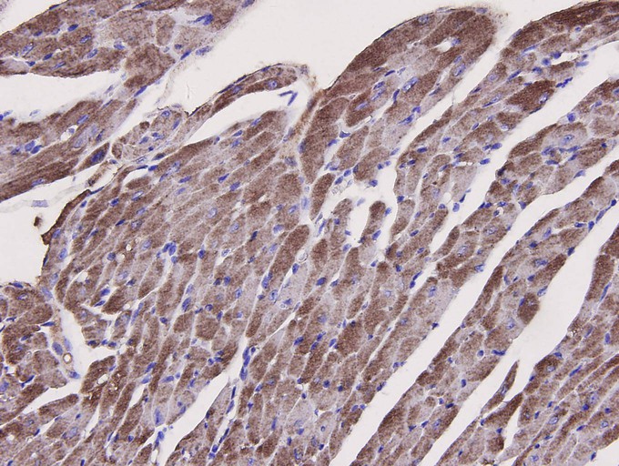
Fig1: Immunohistochemical analysis of paraffin-embedded mouse heart tissue using anti-Caspase-1 antibody. The section was pre-treated using heat mediated antigen retrieval with Tris-EDTA buffer (pH 8.0-8.4) for 20 minutes.The tissues were blocked in 5% BSA for 30 minutes at room temperature, washed with ddH2O and PBS, and then probed with the primary antibody (M1505-2, 1/200) for 30 minutes at room temperature. The detection was performed using an HRP conjugated compact polymer system. DAB was used as the chromogen. Tissues were counterstained with hematoxylin and mounted with DPX. |
|
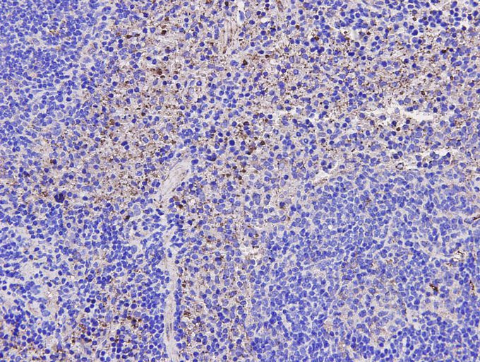
Fig2: Immunohistochemical analysis of paraffin-embedded mouse spleen tissue using anti-Caspase-1 antibody. The section was pre-treated using heat mediated antigen retrieval with Tris-EDTA buffer (pH 8.0-8.4) for 20 minutes.The tissues were blocked in 5% BSA for 30 minutes at room temperature, washed with ddH2O and PBS, and then probed with the primary antibody (M1505-2, 1/200) for 30 minutes at room temperature. The detection was performed using an HRP conjugated compact polymer system. DAB was used as the chromogen. Tissues were counterstained with hematoxylin and mounted with DPX. |
|
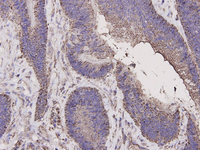
Fig3: Immunohistochemical analysis of paraffin-embedded human colon carcinoma tissue using anti-Caspase-1 antibody. The section was pre-treated using heat mediated antigen retrieval with Tris-EDTA buffer (pH 8.0-8.4) for 20 minutes.The tissues were blocked in 5% BSA for 30 minutes at room temperature, washed with ddH2O and PBS, and then probed with the primary antibody (M1505-2, 1/200) for 30 minutes at room temperature. The detection was performed using an HRP conjugated compact polymer system. DAB was used as the chromogen. Tissues were counterstained with hematoxylin and mounted with DPX. |
|
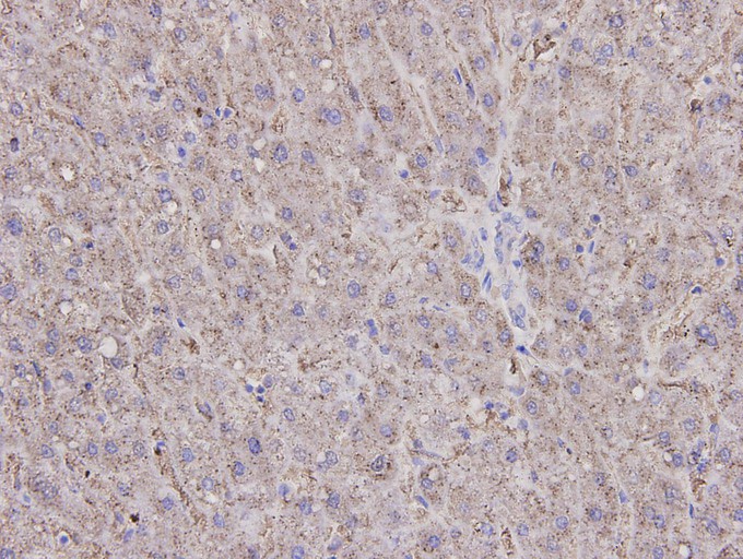
Fig4: Immunohistochemical analysis of paraffin-embedded human liver tissue using anti-Caspase-1 antibody. The section was pre-treated using heat mediated antigen retrieval with Tris-EDTA buffer (pH 8.0-8.4) for 20 minutes.The tissues were blocked in 5% BSA for 30 minutes at room temperature, washed with ddH2O and PBS, and then probed with the primary antibody (M1505-2, 1/200) for 30 minutes at room temperature. The detection was performed using an HRP conjugated compact polymer system. DAB was used as the chromogen. Tissues were counterstained with hematoxylin and mounted with DPX. |
|
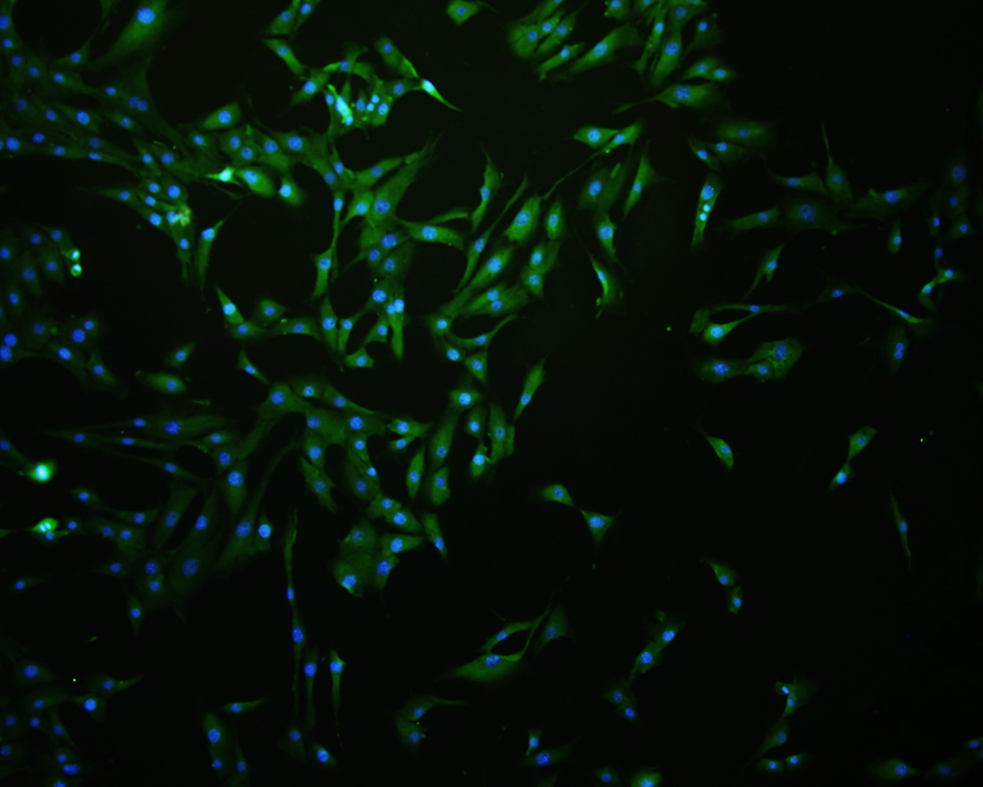
Fig5: ICC staining of Caspase-1 in NIH/3T3 cells (green). Formalin fixed cells were permeabilized with 0.1% Triton X-100 in TBS for 10 minutes at room temperature and blocked with 1% Blocker BSA for 15 minutes at room temperature. Cells were probed with the primary antibody (M1505-2, 1/200) for 1 hour at room temperature, washed with PBS. Alexa Fluor®488 Goat anti-Rabbit IgG was used as the secondary antibody at 1/1,000 dilution. The nuclear counter stain is DAPI (blue). |
Note: All products are “FOR RESEARCH USE ONLY AND ARE NOT INTENDED FOR DIAGNOSTIC OR THERAPEUTIC USE”.