Myc tag Rabbit Polyclonal Antibody

cat.: R1208-1
| Product Type: | Rabbit polyclonal IgG, primary antibodies |
|---|---|
| Species reactivity: | Species independent |
| Applications: | WB, IP, IF-Cell, FC |
| Clonality: | Polyclonal |
| Form: | Liquid |
| Storage condition: | Shipped at 4℃. Store at +4℃ short term (1-2 weeks). Store at -20℃ long term. |
| Storage buffer: | 1*PBS (pH7.4), 0.2% BSA, 40% Glycerol. Preservative: 0.05% Sodium Azide. |
| Concentration: | 1ug/ul |
| Purification: | Immunogen affinity purified. |
| Isotype: | IgG |
| Immunogen: | Synthetic peptide within human Myc aa 410-420. |
| Positive control: | PG-CM cell lysates, C-terminal Myc-tagged recombinant protein, N-terminal Myc-tagged recombinant protein. |
| Recommended Dilutions:
WB IP IF-Cell FC |
1:20,000-1:50,000 2-5 µg/ml. 1:200 1:1,000 |
| Uniprot #: | SwissProt: P01106 Human |
| Alternative names: | avian myelocytomatosis viral oncogene homolog bHLHe39 c-Myc class E basic helix-loop-helix protein 39 MRTL MYC Myc Epitope Tag myc proto-oncogene protein myc-related translation/localization regulatory factor oncogene c-Myc proto-oncogene c-Myc protooncogene homologous to myelocytomatosis virus transcription factor p64 v-myc avian myelocytomatosis viral oncogene homolog v-myc myelocytomatosis viral oncogene homolog (avian) |
Images

|
Fig1:
Western blot analysis of Myc tag on different lysates with Rabbit anti-Myc tag antibody (R1208-1) at 1/20,000 dilution. Lane 1: 293T cell lysate Lane 2: 293T transfected with Myc-tagged Claudin18.2 (C-terminal) cell lysate Lane 3: 293T transfected with Myc-tagged Histone H3.1 (N-terminal) cell lysate Lysates/proteins at 10 µg/Lane. Exposure time: 2 seconds; ECL: K1801; 4-20% SDS-PAGE gel. Proteins were transferred to a PVDF membrane and blocked with 5% NFDM/TBST for 1 hour at room temperature. The primary antibody (R1208-1) at 1/20,000 dilution was used in 5% NFDM/TBST at 4℃ overnight. Goat Anti-Rabbit IgG - HRP Secondary Antibody (HA1001) at 1/50,000 dilution was used for 1 hour at room temperature. |
|
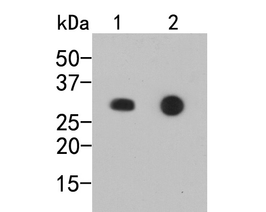
Fig2:
Western blot analysis of Myc tag on different lysates. Proteins were transferred to a PVDF membrane and blocked with 5% BSA in PBS for 1 hour at room temperature. The primary antibody (R1208-1, 1/1,000) was used in 5% BSA at room temperature for 2 hours. Goat Anti-Rabbit IgG - HRP Secondary Antibody (HA1001) at 1:5,000 dilution was used for 1 hour at room temperature. Positive control: Lane 1: C-terminal Myc-tagged recombinant protein Lane 2: N-terminal Myc-tagged recombinant protein |

|
Fig3:
Myc tag was immunoprecipitated in 2µg C terminal Myc Tag fusion protein lysate with R1208-1 at 2 µg/20 µl agarose. Western blot was performed from the immunoprecipitate using EM31105 at 1/1000 dilution. Anti-Mouse IgG - HRP Secondary Antibody (HA1006) at 1:20,000 dilution was used for 60 mins at room temperature. Lane 1: Myc Tag fusion protein lysate (input). Lane 2: R1208-1 IP in Myc Tag fusion protein lysate. Lane 3: Rabbit IgG instead of R1208-1 in Myc Tag fusion protein lysate. Blocking/Dilution buffer: 5% NFDM/TBST |

|
Fig4:
Myc tag was immunoprecipitated in 2µg N terminal Myc Tag fusion protein lysate with R1208-1 at 2 µg/20 µl agarose. Western blot was performed from the immunoprecipitate using EM31105 at 1/1000 dilution. Anti-Mouse IgG - HRP Secondary Antibody (HA1006) at 1:20,000 dilution was used for 60 mins at room temperature. Lane 1: Myc Tag fusion protein lysate (input). Lane 2: R1208-1 IP in Myc Tag fusion protein lysate. Lane 3: Rabbit IgG instead of R1208-1 in Myc Tag fusion protein lysate. Blocking/Dilution buffer: 5% NFDM/TBST |

|
Fig5:
Immunocytochemistry analysis of 293T cells labeling Myc tag with Rabbit anti-Myc tag antibody (R1208-1) at 1/200 dilution. 293T cells, transfected with Myc-tagged empty control, Claudin18.2 (C-terminal) or Histone H3.1 (N-terminal) expression vector, respectively, were fixed in 4% paraformaldehyde for 10 minutes at room temperature, permeabilized with 0.1% Triton X-100 in PBS for 15 minutes at room temperature, then blocked with 1% BSA in 10% negative goat serum for 1 hour at room temperature. Cells were then incubated with Rabbit anti-Myc tag antibody (R1208-1) at 1/200 dilution in 1% BSA in PBST overnight at 4 ℃. Goat Anti-Rabbit IgG H&L (iFluor™ 594, HA1122) was used as the secondary antibody at 1/1,000 dilution. PBS instead of the primary antibody was used as the secondary antibody only control. Nuclear DNA was labelled in blue with DAPI. Myc Tag (HA601081, green) was stained at 1/1,000 dilution overnight at +4℃. Goat Anti-Mouse IgG H&L (iFluor™ 488, HA1125) was used as the secondary antibody at 1/1,000 dilution. |

|
Fig6:
Flow cytometric analysis of 293T cells transfected C-myc-tag labeling Myc tag. Cells were fixed and permeabilized. Then stained with the primary antibody (R1208-1, 1μg/mL) (red) compared with Rabbit IgG Isotype Control (green). After incubation of the primary antibody at +4℃ for an hour, the cells were stained with a iFluor™ 488 conjugate-Goat anti-Rabbit IgG Secondary antibody (HA1121) at 1/1,000 dilution for 30 minutes at +4℃. Unlabelled sample was used as a control (cells without incubation with primary antibody; black). |
Note: All products are “FOR RESEARCH USE ONLY AND ARE NOT INTENDED FOR DIAGNOSTIC OR THERAPEUTIC USE”.