Beclin 1 Rabbit Polyclonal Antibody

cat.: R1509-1
| Product Type: | Rabbit polyclonal IgG, primary antibodies |
|---|---|
| Species reactivity: | Human, Mouse, Rat |
| Applications: | WB, IF-Cell, IF-Tissue, IHC-P |
| Clonality: | Polyclonal |
| Form: | Liquid |
| Storage condition: | Store at +4℃ after thawing. Aliquot store at -20℃. Avoid repeated freeze / thaw cycles. |
| Storage buffer: | 1*PBS (pH7.4), 0.2% BSA, 40% Glycerol. Preservative: 0.05% Sodium Azide. |
| Concentration: | 1ug/ul |
| Purification: | Immunogen affinity purified. |
| Molecular weight: | Predicted band size: 52 kDa |
| Isotype: | IgG |
| Immunogen: | Synthetic peptide within C-terminal human Beclin 1. |
| Positive control: | Jurkat cell lysate, 293 cell lysate, human liver carcinoma tissue, mouse brain tissue, mouse kidney tissue, AGS, Hela, Rat spinal cord tissue lysates. |
| Subcellular location: | Endoplasmic reticulum membrane. |
| Recommended Dilutions:
WB IHC-P IF-Cell IF-Tissue |
1:500-1:2,000 1:200 1:50-1:100 1:50-1:100 |
| Uniprot #: | SwissProt: Q14457 Human | O88597 Mouse |
| Alternative names: | APG6 ATG 6 ATG6 ATG6 autophagy related 6 homolog Bcl-2-interacting protein beclin Beclin 1 (coiled coil moesin like BCL2 interacting protein) Beclin 1 autophagy related Beclin-1 Beclin1 BECN 1 Becn1 BECN1_HUMAN Coiled coil myosin like BCL2 interacting protein Coiled-coil myosin-like BCL2-interacting protein GT197 Protein GT197 VPS 30 VPS30 |
Images
|
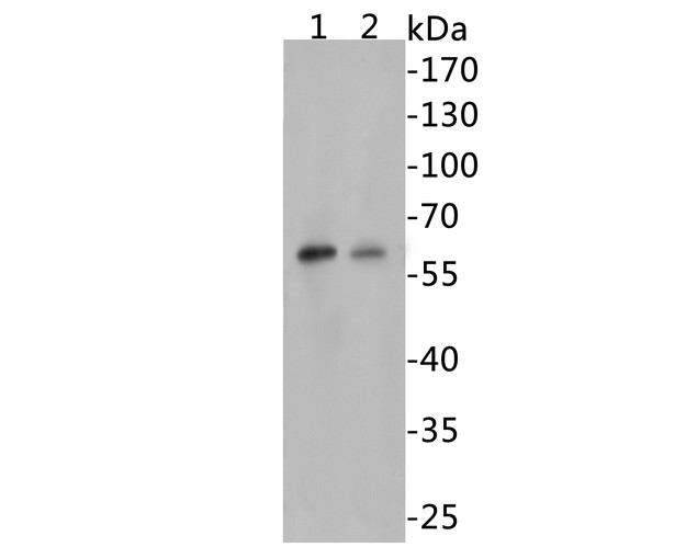
Fig1:
Western blot analysis of Beclin 1 on different lysates. Proteins were transferred to a PVDF membrane and blocked with 5% BSA in PBS for 1 hour at room temperature. The primary antibody (R1509-1, 1/500) was used in 5% BSA at room temperature for 2 hours. Goat Anti-Rabbit IgG - HRP Secondary Antibody (HA1001) at 1:5,000 dilution was used for 1 hour at room temperature. Positive control: Lane 1: Jurkat cell lysates Lane 2: 293 cell lysates |
|
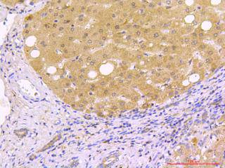
Fig2: Immunohistochemical analysis of paraffin-embedded human liver carcinoma tissue using anti-Beclin 1 antibody. The section was pre-treated using heat mediated antigen retrieval with sodium citrate buffer (pH 6.0) for 20 minutes. The tissues were blocked in 5% BSA for 30 minutes at room temperature, washed with ddH2O and PBS, and then probed with the primary antibody (R1509-1, 1/50) for 30 minutes at room temperature. The detection was performed using an HRP conjugated compact polymer system. DAB was used as the chromogen. Tissues were counterstained with hematoxylin and mounted with DPX. |
|
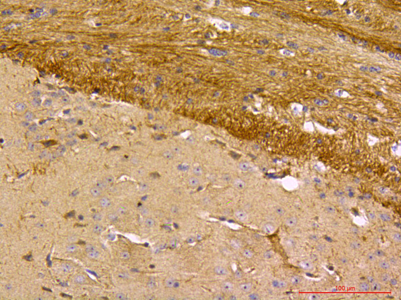
Fig3: Immunohistochemical analysis of paraffin-embedded mouse brain tissue using anti-Beclin 1 antibody. The section was pre-treated using heat mediated antigen retrieval with sodium citrate buffer (pH 6.0) for 20 minutes. The tissues were blocked in 5% BSA for 30 minutes at room temperature, washed with ddH2O and PBS, and then probed with the primary antibody (R1509-1, 1/50) for 30 minutes at room temperature. The detection was performed using an HRP conjugated compact polymer system. DAB was used as the chromogen. Tissues were counterstained with hematoxylin and mounted with DPX. |
|
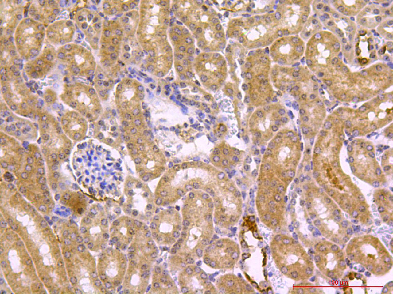
Fig4: Immunohistochemical analysis of paraffin-embedded mouse kidney tissue using anti-Beclin 1 antibody. The section was pre-treated using heat mediated antigen retrieval with sodium citrate buffer (pH 6.0) for 20 minutes. The tissues were blocked in 5% BSA for 30 minutes at room temperature, washed with ddH2O and PBS, and then probed with the primary antibody (R1509-1, 1/50) for 30 minutes at room temperature. The detection was performed using an HRP conjugated compact polymer system. DAB was used as the chromogen. Tissues were counterstained with hematoxylin and mounted with DPX. |
|
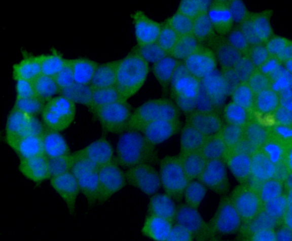
Fig5: ICC staining of Beclin 1 in AGS cells (green). Formalin fixed cells were permeabilized with 0.1% Triton X-100 in TBS for 10 minutes at room temperature and blocked with 1% Blocker BSA for 15 minutes at room temperature. Cells were probed with the primary antibody (R1509-1, 1/100) for 1 hour at room temperature, washed with PBS. Alexa Fluor®488 Goat anti-Rabbit IgG was used as the secondary antibody at 1/1,000 dilution. The nuclear counter stain is DAPI (blue). |
|
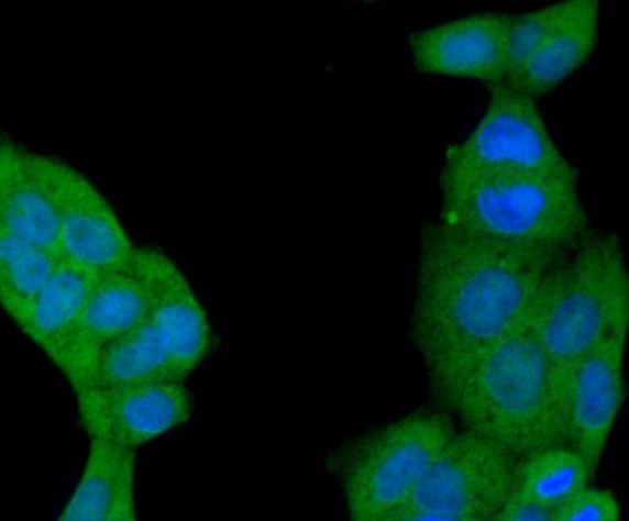
Fig6: ICC staining of Beclin 1 in Hela cells (green). Formalin fixed cells were permeabilized with 0.1% Triton X-100 in TBS for 10 minutes at room temperature and blocked with 1% Blocker BSA for 15 minutes at room temperature. Cells were probed with the primary antibody (R1509-1, 1/100) for 1 hour at room temperature, washed with PBS. Alexa Fluor®488 Goat anti-Rabbit IgG was used as the secondary antibody at 1/1,000 dilution. The nuclear counter stain is DAPI (blue). |
|
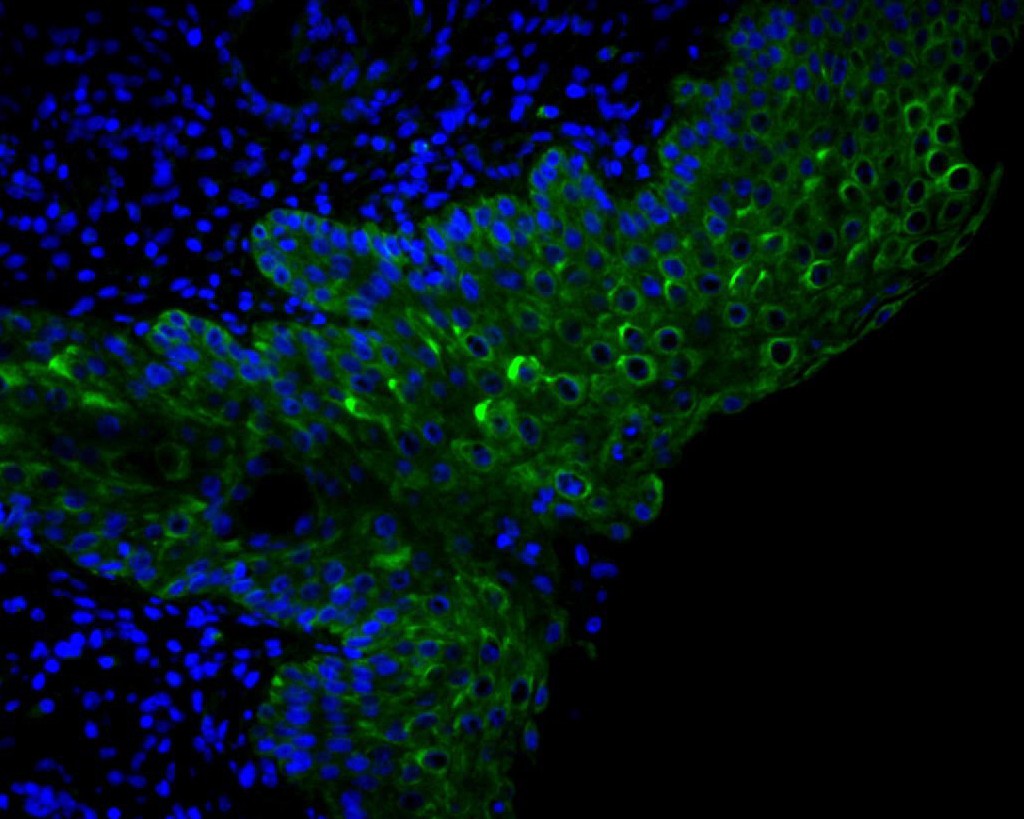
Fig7: IF analysis of paraffin-embedded human liver carcinoma tissue using anti-Beclin 1 antibody. The section was pre-treated using heat mediated antigen retrieval with sodium citrate buffer (pH 6.0) for 20 minutes. The tissues were blocked in 5% BSA for 30 minutes at room temperature, washed with ddH2O and PBS, and then probed with the primary antibody (R1509-1, 1/50) for 30 minutes at room temperature. The detection was performed using an FITC conjugated compact polymer system. DAB was used as the chromogen. Tissues were counterstained with hematoxylin and mounted with DPX. |
|
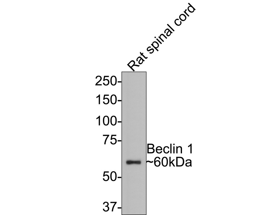
Fig8:
Western blot analysis of Beclin 1 on Rat spinal cord tissue lysates with Rabbit anti-Beclin 1 antibody (R1509-1) at 1/500 dilution. Lysates/proteins at 20 µg/Lane. Predicted band size: 52 kDa Observed band size: 60 kDa Exposure time: 2 minutes; 8% SDS-PAGE gel. Proteins were transferred to a PVDF membrane and blocked with 5% NFDM/TBST for 1 hour at room temperature. The primary antibody (R1509-1) at 1/500 dilution was used in 5% NFDM/TBST at room temperature for 2 hours. Goat Anti-Rabbit IgG - HRP Secondary Antibody (HA1001) at 1:300,000 dilution was used for 1 hour at room temperature. |
Note: All products are “FOR RESEARCH USE ONLY AND ARE NOT INTENDED FOR DIAGNOSTIC OR THERAPEUTIC USE”.