HIF-1 alpha Rabbit Polyclonal Antibody

cat.: R1510-5
| Product Type: | Rabbit polyclonal IgG, primary antibodies |
|---|---|
| Species reactivity: | Human, Mouse, Rat |
| Applications: | WB, IF-Cell, IHC-P, FC |
| Clonality: | Polyclonal |
| Form: | Liquid |
| Storage condition: | Store at +4℃ after thawing. Aliquot store at -20℃ or -80℃. Avoid repeated freeze / thaw cycles. |
| Storage buffer: | 1*PBS (pH7.4), 0.2% BSA, 40% Glycerol. Preservative: 0.05% Sodium Azide. |
| Concentration: | 1ug/ul |
| Purification: | Immunogen affinity purified. |
| Molecular weight: | Predicted band size: 93 kDa |
| Isotype: | IgG |
| Immunogen: | Synthetic peptide within human HIF-1 alpha aa 286-330. |
| Positive control: | Hela, PANC-1, SW480, human tonsil tissue, human colon carcinoma tissue, mouse colon tissue, mouse small intestine tissue, HepG2 cell lysate, A549 cell lysate, Hela cell lysate, PANC-1 cell lysate, SW480 cell lysate, A431 cell lysate, PC-12 cell lysate. |
| Subcellular location: | Cytoplasm, Nucleus, Nucleus speckle |
| Recommended Dilutions:
IF-Cell IHC-P FC WB |
1:50-1:200 1:50-1:200 1:50-1:100 1:500 |
| Uniprot #: | SwissProt: Q16665 Human | Q61221 Mouse | O35800 Rat |
| Alternative names: | ARNT interacting protein ARNT-interacting protein Basic helix loop helix PAS protein MOP1 Basic-helix-loop-helix-PAS protein MOP1 bHLHe78 Class E basic helix-loop-helix protein 78 HIF 1A HIF 1alpha HIF-1-alpha HIF1 A HIF1 Alpha HIF1 HIF1-alpha HIF1A HIF1A_HUMAN Hypoxia inducible factor 1 alpha Hypoxia inducible factor 1 alpha isoform I.3 Hypoxia inducible factor 1 alpha subunit Hypoxia inducible factor 1 alpha subunit basic helix loop helix transcription factor Hypoxia inducible factor 1, alpha subunit (basic helix loop helix transcription factor) Hypoxia inducible factor1alpha Hypoxia-inducible factor 1-alpha Member of PAS protein 1 Member of PAS superfamily 1 Member of the PAS Superfamily 1 MOP 1 MOP1 PAS domain-containing protein 8 PASD 8 PASD8 |
Images
|
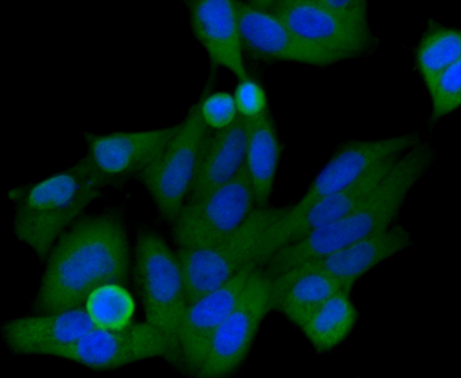
Fig1: Immunocytochemical staining of Hela cells using anti-HIF-1 alpha rabbit polyclonal antibody. . |
|
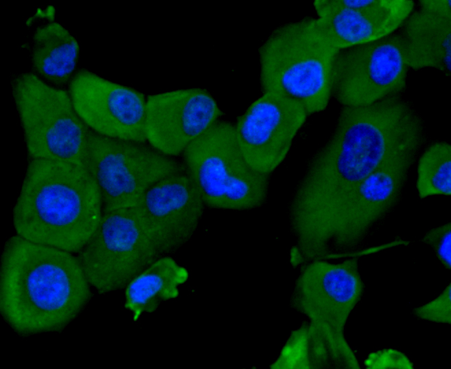
Fig2: Immunocytochemical staining of PANC-1 cells using anti-HIF-1 alpha rabbit polyclonal antibody. . |
|
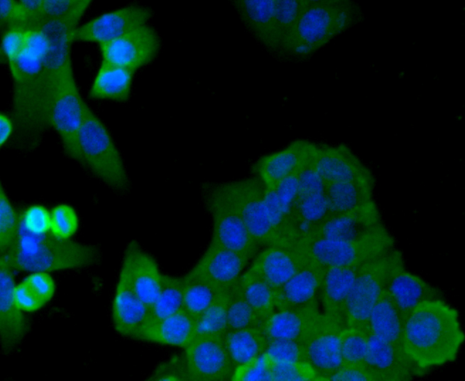
Fig3: Immunocytochemical staining of SW480 cells using anti-HIF-1 alpha rabbit polyclonal antibody. . |
|
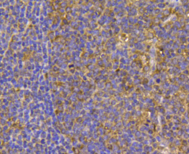
Fig4: Immunohistochemical analysis of paraffin- embedded human tonsil tissue using anti-HIF-1 alpha rabbit polyclonal antibody. . |
|
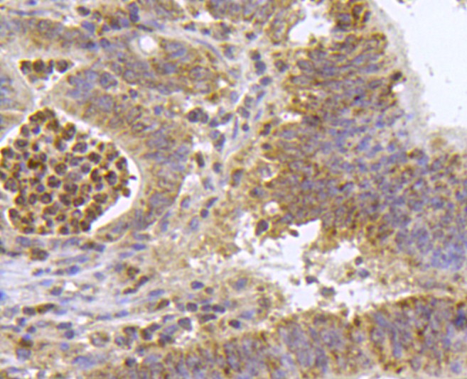
Fig5: Immunohistochemical analysis of paraffin- embedded human colon carcinoma tissue using anti-HIF-1 alpha rabbit polyclonal antibody. . |
|
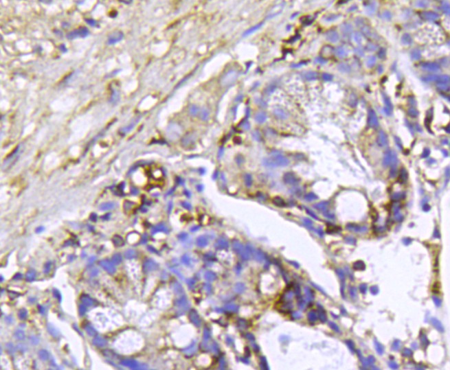
Fig6: Immunohistochemical analysis of paraffin- embedded mouse colon tissue using anti-HIF-1 alpha rabbit polyclonal antibody. . |
|
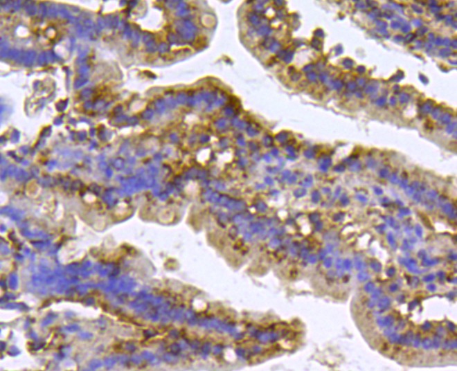
Fig7: Immunohistochemical analysis of paraffin- embedded mouse small intestine tissue using anti-HIF-1 alpha rabbit polyclonal antibody. . |
|
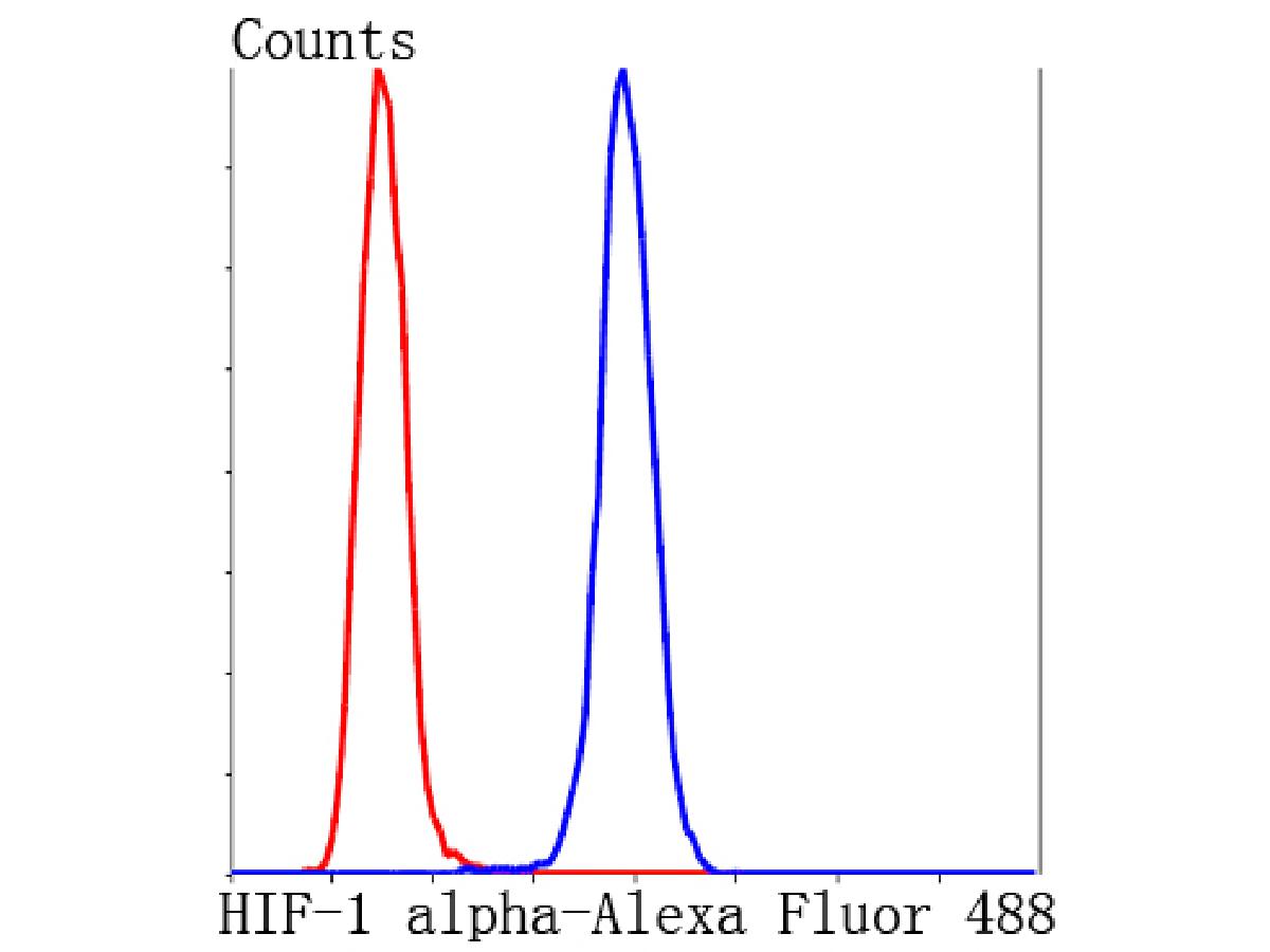
Fig8: Flow cytometric analysis of Hela cells with HIF-1 alpha antibody at 1/100 dilution (blue) compared with an unlabelled control (cells without incubation with primary antibody; red). Alexa Fluor 488-conjugated Goat anti rabbit IgG was used as the secondary antibody. |
|
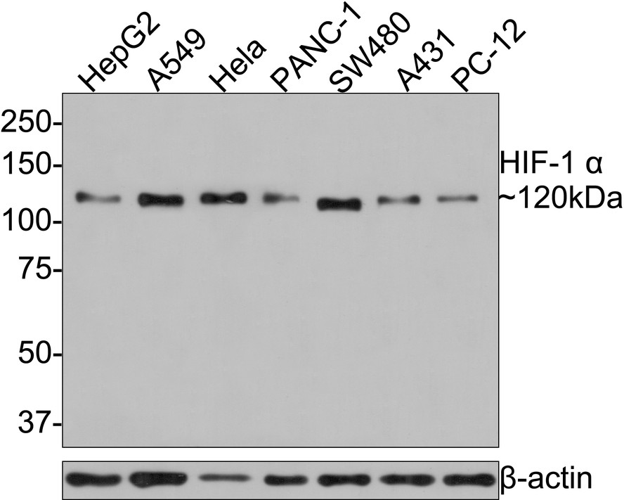
Fig9:
Western blot analysis of HIF-1 alpha on different lysates with Rabbit anti-HIF-1 alpha antibody (R1510-5) at 1/500 dilution. Lane 1: HepG2 cell lysate Lane 2: A549 cell lysate Lane 3: Hela cell lysate Lane 4: PANC-1 cell lysate Lane 5: SW480 cell lysate Lane 6: A431 cell lysate Lane 7: PC-12 cell lysate Lysates/proteins at 10 µg/Lane. Predicted band size: 93 kDa Observed band size: 120 kDa Exposure time: 1 minute; 8% SDS-PAGE gel. Proteins were transferred to a PVDF membrane and blocked with 5% NFDM/TBST for 1 hour at room temperature. The primary antibody (R1510-5) at 1/500 dilution was used in 5% NFDM/TBST at room temperature for 2 hours. Goat Anti-Rabbit IgG - HRP Secondary Antibody (HA1001) at 1:300,000 dilution was used for 1 hour at room temperature. |
Note: All products are “FOR RESEARCH USE ONLY AND ARE NOT INTENDED FOR DIAGNOSTIC OR THERAPEUTIC USE”.