ID2 Rabbit Polyclonal Antibody

cat.: R1510-6
| Product Type: | Rabbit polyclonal IgG, primary antibodies |
|---|---|
| Species reactivity: | Human, Mouse |
| Applications: | WB, IHC-P, FC |
| Clonality: | Polyclonal |
| Form: | Liquid |
| Storage condition: | Store at +4℃ after thawing. Aliquot store at -20℃ or -80℃. Avoid repeated freeze / thaw cycles. |
| Storage buffer: | 1*PBS (pH7.4), 0.2% BSA, 40% Glycerol. Preservative: 0.05% Sodium Azide. |
| Concentration: | 1ug/ul |
| Purification: | Immunogen affinity purified. |
| Molecular weight: | 15 kDa |
| Isotype: | IgG |
| Immunogen: | Synthetic peptide within C-terminal human ID2. |
| Positive control: | NCCIT cell lysates, human fetal skeletal muscle tissue, human kidney tissue, mouse colon tissue, mouse lung tissue, HepG2. |
| Subcellular location: | Cytoplasm, Nucleus. |
| Recommended Dilutions:
WB IHC-P FC |
1:500 1:100-1:500 1:50-1:100 |
| Uniprot #: | SwissProt: Q02363 Human | P41136 Mouse |
| Alternative names: | bHLHb26 Cell growth inhibiting gene 8 class B basic helix loop helix protein 26 Class B basic helix-loop-helix protein 26 DNA binding protein inhibitor ID 2 DNA binding protein inhibitor ID2 DNA-binding protein inhibitor ID-2 GIG 8 GIG8 Helix loop helix protein ID2 ID2 ID2_HUMAN ID2A ID2H Inhibitor of differentiation 2 Inhibitor of DNA binding 2 Inhibitor of DNA binding 2, dominant ne |
Images
|
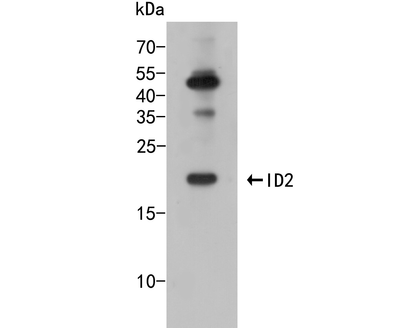
Fig1: Western blot analysis of ID2 on NCCIT cell lysate. Proteins were transferred to a PVDF membrane and blocked with 5% BSA in PBS for 1 hour at room temperature. The primary antibody (R1510-6, 1/500) was used in 5% BSA at room temperature for 2 hours. Goat Anti-Rabbit IgG - HRP Secondary Antibody (HA1001) at 1:5,000 dilution was used for 1 hour at room temperature. |
|
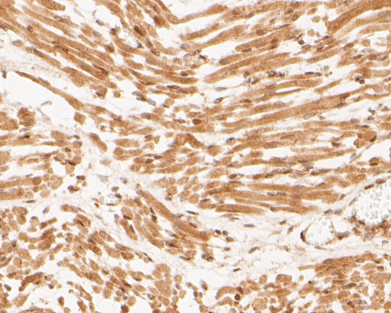
Fig2: Immunohistochemical analysis of paraffin-embedded human fetal skeletal muscle tissue using anti-ID2 antibody. The section was pre-treated using heat mediated antigen retrieval with sodium citrate buffer (pH 6.0) for 20 minutes. The tissues were blocked in 5% BSA for 30 minutes at room temperature, washed with ddH2O and PBS, and then probed with the primary antibody (R1510-6, 1/400) for 30 minutes at room temperature. The detection was performed using an HRP conjugated compact polymer system. DAB was used as the chromogen. Tissues were counterstained with hematoxylin and mounted with DPX. |
|
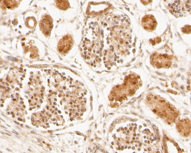
Fig3: Immunohistochemical analysis of paraffin-embedded human kidney tissue using anti-ID2 antibody. The section was pre-treated using heat mediated antigen retrieval with sodium citrate buffer (pH 6.0) for 20 minutes. The tissues were blocked in 5% BSA for 30 minutes at room temperature, washed with ddH2O and PBS, and then probed with the primary antibody (R1510-6, 1/400) for 30 minutes at room temperature. The detection was performed using an HRP conjugated compact polymer system. DAB was used as the chromogen. Tissues were counterstained with hematoxylin and mounted with DPX. |
|
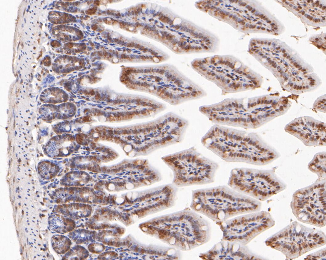
Fig4: Immunohistochemical analysis of paraffin-embedded mouse colon tissue using anti-ID2 antibody. The section was pre-treated using heat mediated antigen retrieval with sodium citrate buffer (pH 6.0) for 20 minutes. The tissues were blocked in 5% BSA for 30 minutes at room temperature, washed with ddH2O and PBS, and then probed with the primary antibody (R1510-6, 1/400) for 30 minutes at room temperature. The detection was performed using an HRP conjugated compact polymer system. DAB was used as the chromogen. Tissues were counterstained with hematoxylin and mounted with DPX. |
|
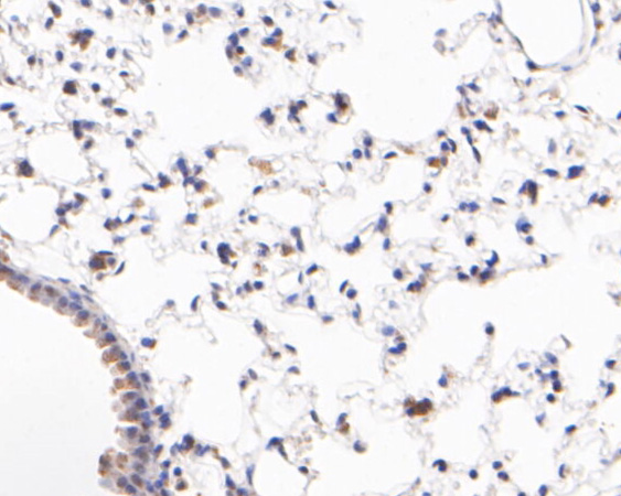
Fig5: Immunohistochemical analysis of paraffin-embedded mouse lung tissue using anti-ID2 antibody. The section was pre-treated using heat mediated antigen retrieval with sodium citrate buffer (pH 6.0) for 20 minutes. The tissues were blocked in 5% BSA for 30 minutes at room temperature, washed with ddH2O and PBS, and then probed with the primary antibody (R1510-6, 1/400) for 30 minutes at room temperature. The detection was performed using an HRP conjugated compact polymer system. DAB was used as the chromogen. Tissues were counterstained with hematoxylin and mounted with DPX. |
|
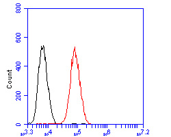
Fig6: Flow cytometric analysis of ID2 was done on HepG2 cells. The cells were fixed, permeabilized and stained with the primary antibody (R1510-6, 1/50) (red). After incubation of the primary antibody at room temperature for an hour, the cells were stained with a Alexa Fluor 488-conjugated Goat anti-Rabbit IgG Secondary antibody at 1/1000 dilution for 30 minutes.Unlabelled sample was used as a control (cells without incubation with primary antibody; black). |
Note: All products are “FOR RESEARCH USE ONLY AND ARE NOT INTENDED FOR DIAGNOSTIC OR THERAPEUTIC USE”.