PD-L1 Rabbit Polyclonal Antibody

cat.: R1511-13
| Product Type: | Rabbit polyclonal IgG, primary antibodies |
|---|---|
| Species reactivity: | Human |
| Applications: | WB, IF-Cell, IHC-P |
| Clonality: | Polyclonal |
| Form: | Liquid |
| Storage condition: | Store at +4℃ after thawing. Aliquot store at -20℃. Avoid repeated freeze / thaw cycles. |
| Storage buffer: | 1*TBS (pH7.4), 0.2% BSA, 50% Glycerol. Preservative: 0.05% Sodium Azide. |
| Concentration: | 1ug/ul |
| Purification: | Protein A affinity purified. |
| Molecular weight: | 33 kDa (Predicted band size) |
| Isotype: | IgG |
| Immunogen: | Recombinant protein within Human CD274 aa 38-272 / 290. |
| Positive control: | MCF-7 cell lysates, MG-63, human lung adenocarcinoma tissue. |
| Subcellular location: | Cell membrane, Endomembrane system. |
| Recommended Dilutions:
WB IF-Cell IHC-P |
1:500-1:1,000 1:50-1:200 1:50-1:200 |
| Uniprot #: | SwissProt: Q9NZQ7 Human |
| Alternative names: | B7 H B7 H1 B7 homolog 1 B7-H1 B7H B7H1 CD 274 CD274 CD274 antigen CD274 molecule MGC142294 MGC142296 OTTHUMP00000021029 PD L1 PD-L1 PD1L1_HUMAN PDCD1 ligand 1 PDCD1L1 PDCD1LG1 PDL 1 PDL1 Programmed cell death 1 ligand 1 Programmed death ligand 1 RGD1566211 |
Images
|
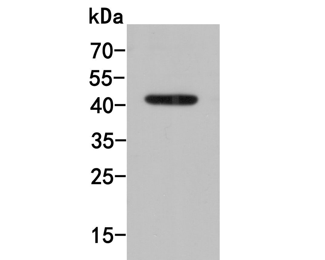
Fig1: Western blot analysis of PD-L1 on MCF-7 cell lysates. Proteins were transferred to a PVDF membrane and blocked with 5% BSA in PBS for 1 hour at room temperature. The primary antibody (R1511-13, 1/500) was used in 5% BSA at room temperature for 2 hours. Goat Anti-Rabbit IgG - HRP Secondary Antibody (HA1001) at 1:5,000 dilution was used for 1 hour at room temperature. |
|
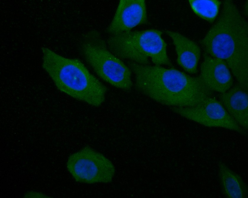
Fig2: ICC staining of PD-L1 in MG-63 cells (green). Formalin fixed cells were permeabilized with 0.1% Triton X-100 in TBS for 10 minutes at room temperature and blocked with 1% Blocker BSA for 15 minutes at room temperature. Cells were probed with the primary antibody (R1511-13, 1/100) for 1 hour at room temperature, washed with PBS. Alexa Fluor®488 Goat anti-Rabbit IgG was used as the secondary antibody at 1/1,000 dilution. The nuclear counter stain is DAPI (blue). |
|
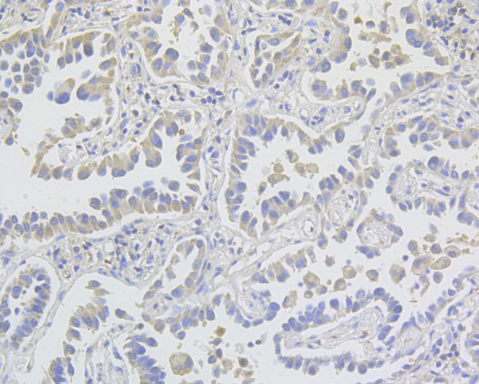
Fig3: Immunohistochemical analysis of paraffin-embedded human lung adenocarcinoma tissue using anti-PD-L1 antibody. The section was pre-treated using heat mediated antigen retrieval with Tris-EDTA buffer (pH 8.0-8.4) for 20 minutes.The tissues were blocked in 5% BSA for 30 minutes at room temperature, washed with ddH2O and PBS, and then probed with the primary antibody (R1511-13, 1/800) for 30 minutes at room temperature. The detection was performed using an HRP conjugated compact polymer system. DAB was used as the chromogen. Tissues were counterstained with hematoxylin and mounted with DPX. |
Note: All products are “FOR RESEARCH USE ONLY AND ARE NOT INTENDED FOR DIAGNOSTIC OR THERAPEUTIC USE”.